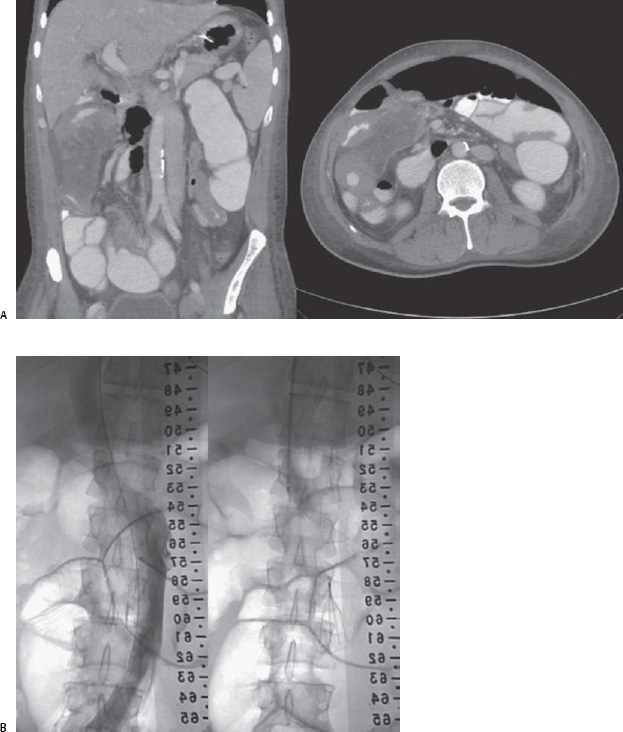
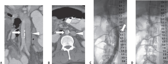
Case 96 A 46-year-old man with deep venous thrombosis (DVT) is scheduled for surgical resection of tumor in his right leg, pelvis, and abdomen. (A,B) Coronal and axial contrast-enhanced computed tomographic (CT) images show transposition of the calcified aorta (arrow) and the inferior vena cava (IVC: arrowhead). (C,D) Venogram and fluoroscopic image after placement of a Tulip filter (Cook Medical, Bloomington, IN). The left-sided IVC drains into the left renal vein (arrow).
 Clinical Presentation
 Imaging Findings
 Differential Diagnosis
 Essential Facts
Stay updated, free articles. Join our Telegram channel

Full access? Get Clinical Tree


