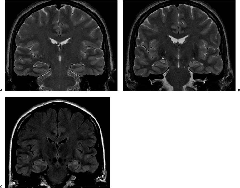
Case 97 A 25-year-old with epilepsy. (A,B) Coronal T2-weighted images (WIs) perpendicular to the long axis of the hippocampus show decreased height of the left hippocampus in comparison with the right and mild architectural distortion (arrows). (C) Fluid-attenuated inversion recovery coronal image at the level of the hippocampal head shows increased signal on the left in comparison with the right and with the remainder of the temporal cortex (arrow). • Mesial temporal sclerosis (MTS): Atrophy of one or both hippocampal formations is indicative of MTS. There is increased T2 signal and architectural distortion. The abnormalities may involve other areas of the temporal lobe. • Herpes encephalitis: This is the most common type of sporadic viral encephalitis, with a predilection for the temporal lobes. Magnetic resonance imaging (MRI) shows T2 hyperintensity corresponding to edematous changes in the temporal lobes, inferior frontal lobes, and insula, with a predilection for the medial temporal lobes. Foci of hemorrhage occasionally can be observed on MRI.
Clinical Presentation
Imaging Findings
Differential Diagnosis
Stay updated, free articles. Join our Telegram channel

Full access? Get Clinical Tree


