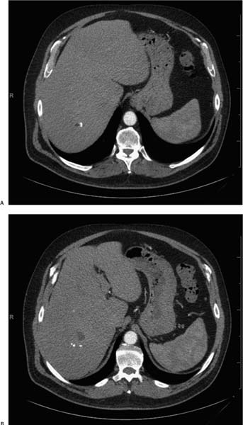
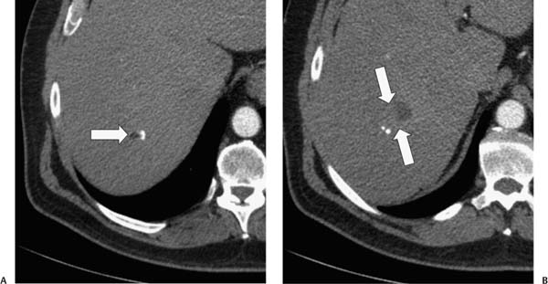
Case 97 A 50-year-old man presents for computed tomography work-up of a liver lesion noted on ultrasound. (A) Contrast-enhanced computed tomography (CT) in the arterial phase shows a focus of macroscopic fat within the right lobe of the liver with an adjacent dense calcification (arrow). (B) More caudal slice shows a heterogeneous soft-tissue mass (arrows) with peripheral dense calcifications. • Metastatic germ cell tumor: This is the principal diagnostic consideration for a mass in the liver with patchy macroscopic fat, a soft-tissue component, and dense calcifications. The calcification in Figure A has the appearance of a tooth.
 Clinical Presentation
 Imaging Findings
 Differential Diagnosis
Stay updated, free articles. Join our Telegram channel

Full access? Get Clinical Tree


