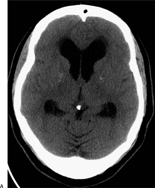
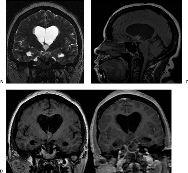
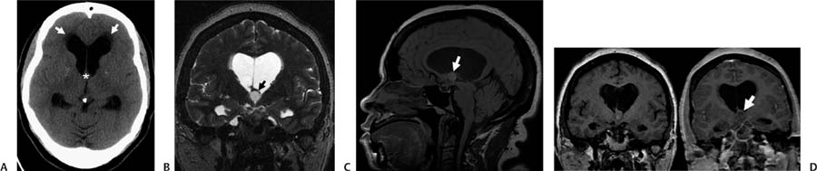
Case 98 A 39-year-old woman with intermittent positional headache. (A) Axial computed tomography (CT) demonstrates enlarged frontal horns and transependymal fluid leak (white arrows), which is indicative of hydrocephalus. There is a mass with attenuation similar to that of white matter in the vicinity of the foramen of Monro (asterisk). (B) Coronal T2-weighted image (WI) shows a mass in the superior aspect of the 3rd ventricle (arrow), with a central intermediate signal and a thin rim of low signal. (C) On the sagittal T1WI, the lesion has increased signal intensity (arrow). The lateral ventricles are dilated, whereas the 3rd and 4th are not. (D) Coronal T1WIs before and after contrast show no enhancement of the mass (arrow).
Clinical Presentation
Further Work-up
Imaging Findings
Differential Diagnosis
Stay updated, free articles. Join our Telegram channel

Full access? Get Clinical Tree


