 Clinical Presentation
An 18-year-old man with left-sided pleuritic chest pain.
Further Work-up
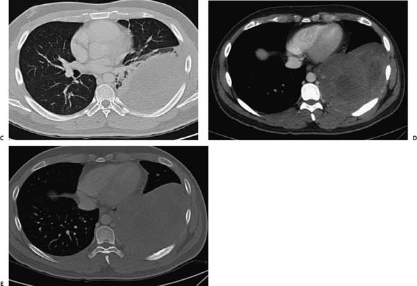
 Imaging Findings

(A) Posteroanterior chest radiograph demonstrates a large mass in the left hemithorax (white arrow). The diaphragmatic silhouette is lost, although a portion of the left border of the heart is still seen, suggesting a posterobasal location (black arrow). (B) Lateral chest radiograph confirms the posterior location of the mass (arrows
Stay updated, free articles. Join our Telegram channel

Full access? Get Clinical Tree


