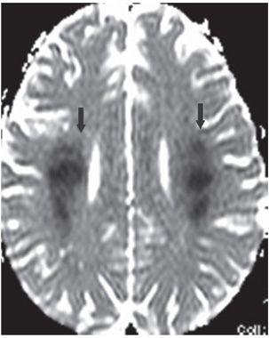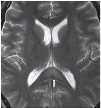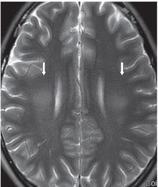


FINDINGS Figure 98-1. Axial ADC map through the splenium of corpus callosum. There is focal restricted diffusion of the splenium (arrow). Figure 98-2. Axial ADC map through the corona radiata. There are bilateral symmetrical white matter (WM) areas of restricted diffusion (arrows). Figures 98-3 and 98-4. Axial T2WI through the splenium and the corona radiata, respectively. There are confluent hyperintensity in the splenium and bilateral WM (arrows).
DIFFERENTIAL DIAGNOSIS Toxic encephalopathy, methotrexate leukoencephalopathy (MTX LE), posterior reversible encephalopathy syndrome (PRES), delayed post-hypoxic leukoencephalopathy (DPHL) hypoglycemia, hypoxic ischemic encephalopathy (HIE).
DIAGNOSIS MTX LE.
DISCUSSION
Stay updated, free articles. Join our Telegram channel

Full access? Get Clinical Tree


