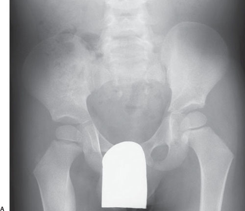
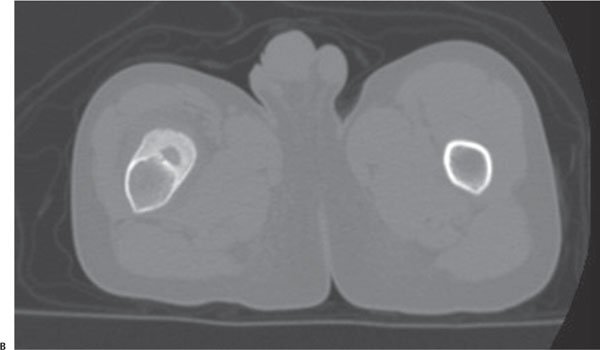
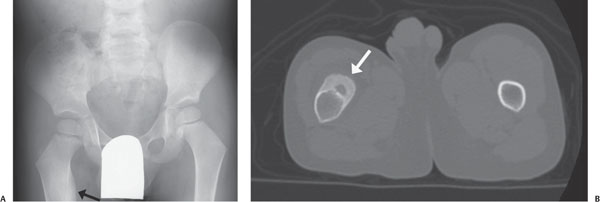
Case 98 A 6-year-old boy with hip pain. (A) Anteroposterior radiograph of the pelvis: there is sclerosis of the proximal diaphysis of the right femur that is incompletely imaged (arrow). (B) Axial computed tomography (CT) image: there is a central, well-defined lytic lesion with thick subperiosteal new bone formation (arrow). • Osteoid osteoma: These findings are consistent with oste-oid osteoma. • Osteomyelitis (Brodie abscess): This can be difficult to differentiate from osteoid osteoma. Often, there is a lucent tract extending from the nidus, as well as cortical destruc-tion. • Stress fracture:
Clinical Presentation
Further Work-up
Imaging Findings
Differential Diagnosis
![]()
Stay updated, free articles. Join our Telegram channel

Full access? Get Clinical Tree


