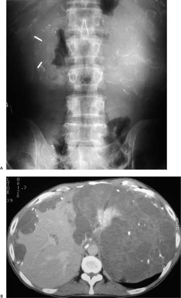
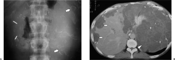
Case 98 A 63-year-old woman presents with abdominal distension and pain. (A) Frontal abdominal radiograph shows curvilinear calcifications throughout the abdomen (arrows). (B) Contrast-enhanced computed tomography in the venous phase shows scalloping of the liver margin (arrows) and nonenhancing, low-density material throughout the abdomen (arrowhead) with septations and scattered calcifications. • Pseudomyxoma peritonei:
 Clinical Presentation
 Imaging Findings
 Differential Diagnosis
![]()
Stay updated, free articles. Join our Telegram channel

Full access? Get Clinical Tree


