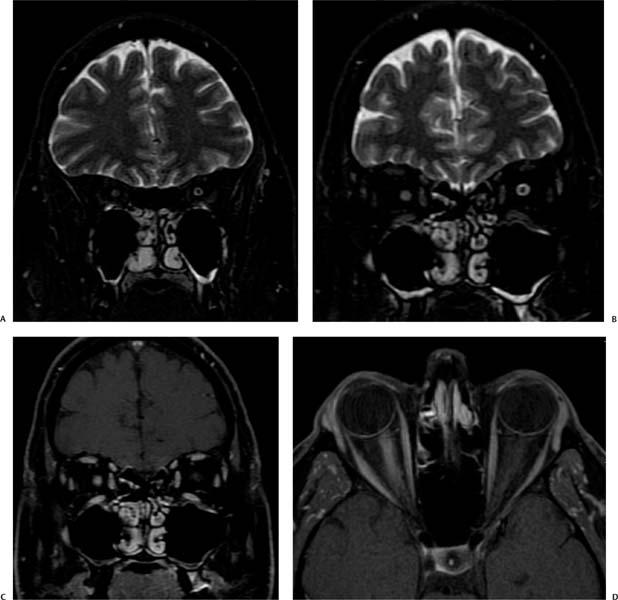
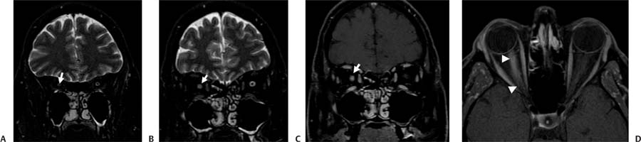
Case 99 A 42-year-old woman presenting with unilateral pain, decreased visual acuity, and loss of color vision. (A,B) Coronal T2-weighted images (WIs) of the orbits show enlargement of the right optic nerve (arrow), resulting in effacement of the right optic nerve sheath in comparison with the left. (C) Coronal postcontrast T1WI shows that the right optic nerve is enlarged and enhances homogeneously (arrow). (D) On the axial T1WI, note how the enhancement extends from the apex of the orbit to the globe (arrowheads). • Optic neuritis: This is inflammatory optic neuropathy resulting in vision loss. Magnetic resonance imaging (MRI) findings may be normal or demonstrate T2 hyperintensity with a normal or mildly enlarged optic nerve. Contrast enhancement can involve a segment or the entire optic nerve. • Sarcoidosis:
Clinical Presentation
Imaging Findings
Differential Diagnosis
![]()
Stay updated, free articles. Join our Telegram channel

Full access? Get Clinical Tree


