 Clinical Presentation
A 52-year-old woman with fever 3 weeks after cardiac valve replacement.
Further Work-up
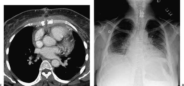
 Imaging Findings
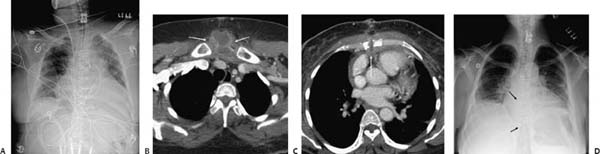
(A) Frontal chest radiograph demonstrates displacement of multiple sternal wires. The lungs are hypoinflated, and linear atelectasis is seen at the lung bases. (B) Contrast-enhanced computed tomography (CT) of the chest (soft-tissue windows) through the upper sternum demonstrates a sternal abscess (arrows). (C) Contrast-enhanced CT of the chest (soft-tissue windows) through the lower sternum shows sternal nonunion with extension of the abscess into the anterior mediastinum. Note the fat stranding and pericardial effusion (arrow
Stay updated, free articles. Join our Telegram channel

Full access? Get Clinical Tree


