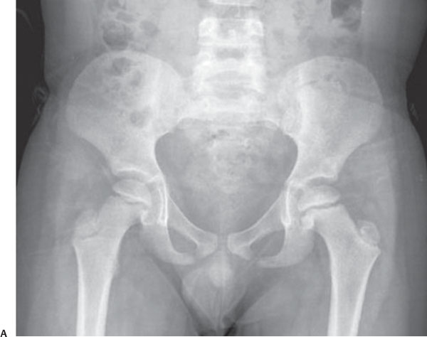
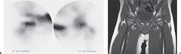
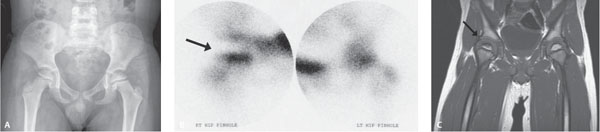
Case 99 A 5-year-old with a limp. (A) Plain film: Both hips appear normal. There is no sclerosis, flattening, or fragmentation. (B) Nuclear medicine methylene diphosphonate (MDP) bone scan of both hips: On the pinhole views, there is decreased activity in the right femoral head (arrow). (C) Magnetic resonance imaging (MRI): T1 shows a hypointense, irregular area in the periphery of the right femoral head (arrow). • Legg-Calvé-Perthes disease (avascular necrosis [AVN]):
Clinical Presentation
Further Work-up
Imaging Findings
Differential Diagnosis
![]()
Stay updated, free articles. Join our Telegram channel

Full access? Get Clinical Tree


