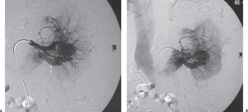
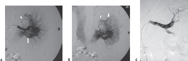
Case 99 The patient is a 59-year-old man who presented with left flank pain. (A) Selected renal angiogram shows early opacification of the renal vein (arrow), dense neovascularity, and an ectatic capsular branch (arrowhead). (B) Delayed image shows renal nephrogram (arrow) and blush within an exophytic mass (arrowhead). (C) Capsular and parenchymal branches were embolized with Gelfoam and particles, and the anterior and posterior divisions of the renal artery were embolized with coils. The patient underwent nephrectomy the following day.
 Clinical Presentation
 Imaging Findings
 Differential Diagnosis
Stay updated, free articles. Join our Telegram channel

Full access? Get Clinical Tree


