Case 99
Case History
A 64-year-old woman, 6 months after right breast lumpectomy and radiation therapy, has new calcifications adjacent to her lumpectomy site.
Physical Examination
• right breast: scar in upper outer quadrant of right breast from previous lumpectomy; no new masses
• left breast: normal exam
Mammogram
Calcifications (Fig. 99–1)
• type: pleomorphic/heterogeneous
• distribution: grouped/clustered
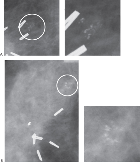
Figure 99–1. In the right upper outer quadrant there are clips associated with architectural distortion from previous lumpectomy. Adjacent to the clips is a group of new heterogeneous calcifications (circled). (A). Right MLO magnification mammogram. (B). Right CC magnification mammogram.
Ultrasound


Stay updated, free articles. Join our Telegram channel

Full access? Get Clinical Tree


