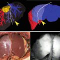
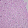
Fig. 25.1
Absorption imaging. Sentinel nodes and lymph vessel were clearly observed in the absorption imaging (b); however, they are not visualized in the ordinary light (a)
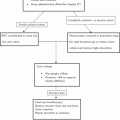
We developed a new IRLS system (OLYMPUS, Tokyo, Japan) for detecting SN using both the absorption and the fluorescence imaging of ICG (Fig. 25.2). One of the characteristics is instant switching the observation mode between the light absorption and the fluorescence imaging. In this study, we determined the feasibility of ICG plus a new infrared ray laparoscopy system (n-IRLS) with dual imaging for SN of gastric cancer.
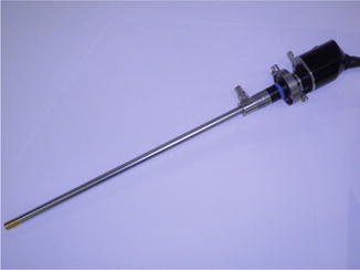

Fig. 25.2
New IRLS system. We developed a new IRLS system (OLYMPUS, Tokyo, Japan) for the detection of SN using both the absorption and the fluorescence imaging of ICG. One of the characteristics is instant switching of the observation mode between the absorption and the fluorescence imaging
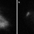
The following are the details of the principle of ICG and infrared ray: It is known that ICG has a maximum absorption of 805 nm wavelength when ICG is combined with plasma protein. In the irradiated light near the maximum absorption wavelength, the ICG area absorbs the light and becomes darker, and other areas without ICG as the background become brighter. This is the mechanism of the infrared light absorption imaging with ICG. On the other hand, ICG in itself emits a fluorescence which has a maximum of 830 nm wavelength. Compared to the reflected light, the intensity of the fluorescence is extremely weak. Thus, infrared fluorescence imaging with ICG is enabled by completely cutting the reflected light and receiving the light near the maximum fluorescence wavelength. With regards to the fluorescence observation, the difference from the above mentioned system is the changed spectral transmission wavelength of the infrared filter in the light source unit from the maximum light absorption wavelength (805 nm) to a shorter wavelength (690–790 nm). The light transmitted through the filter passes through the light guide and the laparoscope to irradiate the subject. Then ICG is excited and emits fluorescence. Inside the camera head, a special filter is used. The filter cuts the light (690–790 nm) reflected from the subject and allows only fluorescence (800 nm or longer) to pass, enabling fluorescence observation. During the absorption the light emitted from the light source unit has the same wavelength as that of the fluorescence. Also by collecting the reflected light at the camera head an absorption image is formed. The wavelength of the light for the absorption observation is changed from the maximum absorption wavelength (805 nm) to a shorter wavelength. However, the absorption images have practically no adverse effect for identifying the SN before clinical use (data not shown).
Materials and Methods
Patients
The study was approved by the Ethics Committee for Biomedical Research of the Jikei Institutional Review Board, and all patients provided informed consent. Patients admitted to the Jikei University Hospital with cT1 gastric cancer without obvious metastases were included prospectively in this study, which was diagnosed by preoperative abdominal CT scans and endoscopic ultrasonography. From February 2012 to December 2013, 13 patients were registered. Table 25.1 shows the patients characteristics and preoperative findings for SNNS for patients with gastric cancer.
Table 25.1
Patients characteristics and preoperative findings for sentinel navigation surgery
Patients characteristics | Preoperative diagnosis | |||||||||
|---|---|---|---|---|---|---|---|---|---|---|
Case | Age (Y) | Gender | Height (cm) | BW (kg) | BMI | Type | Size (mm) | Location | Depth | Pathology |
1 | 60 | M | 170 | 73 | 25.26 | 0–IIc | 10 | M/less | M | Poor |
2 | 77 | F | 145 | 79 | 37.57 | 0–IIc | 10 | M/less | M | Poor |
3 | 57 | M | 177 | 66 | 21.07 | 0–IIc | 10 | U | SM | Well |
4 | 74 | M | 166 | 51 | 18.5 | 0–IIc | 30 | M | SM | Well |
5 | 78 | M
Stay updated, free articles. Join our Telegram channel
Full access? Get Clinical Tree
 Get Clinical Tree app for offline access

| ||||||||
