The abdomen consists of four regions:
Anterior abdominal wall
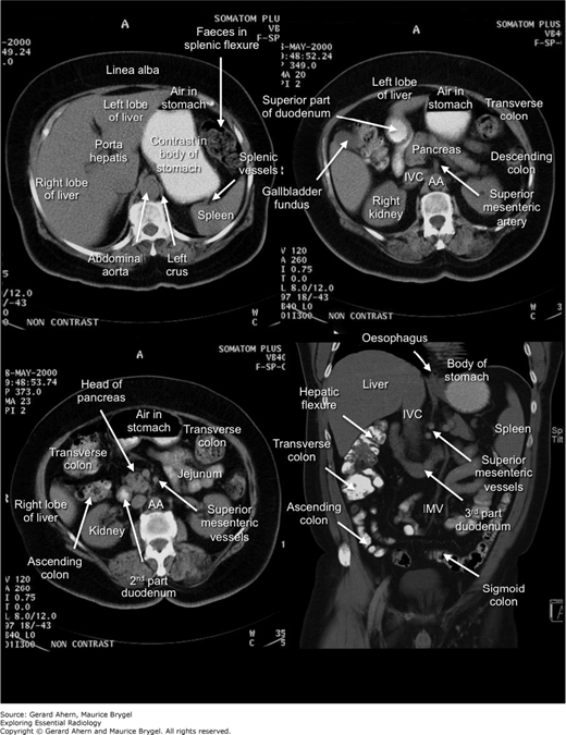
Abdominal cavity (with viscera)
Inguinal canal
Posterior abdominal wall
Imaging of the anterior abdominal wall seldom shows significant pathology though certain conditions may be seen on ultrasound, such as a Spigelian hernia.
The abdominal cavity, with all its various viscera, is a rich source of pathological images.
The posterior abdominal wall contains the major vessels. The kidneys and ureters have been included as abdominal viscera, though they are really constituents of the posterior abdominal wall.


Epigastric pain, indigestion, nausea, and cachexia.
Focal mass lesion on greater curvature of stomach (gastric carcinoma).
Resection if not too late.

Cachexia, constipation, diarrhea, and peri-rectal bleeding.
Gastrografin enema reveals stenotic lesion in proximal transverse colon similar to an ‘apple core’ (colonic carcinoma).
Resection and anastomosis.

History of cause (hernia, adhesions) with vomiting, colicky pain, distension, and constipation.
Gas-filled distended loops of small bowel (note circular folds) with lack of gas in the large bowel. Mechanical small bowel obstruction.
Treat cause; nasogastric suction; intravenous fluids; analgesia.

History of cause (hernia, adhesions) with vomiting, colicky pain, distension, and constipation.
Gas-filled distended loops of small bowel with fluid levels. Mechanical small bowel obstruction.
Treat cause; nasogastric suction; intravenous fluids; analgesia.

History of cause (volvulus, constipation, tumor, congenital) with colicky pain, distension, and constipation.
Dilated large bowel with obvious haustra and peripheral location. Large bowel obstruction.
Treat cause and relieve obstruction. Usually caused by carcinoma.

History of cause (volvulus, constipation, tumor, congenital) with colicky pain, distension and constipation.
Air-fluid level seen in dilated caecum and ascending colon (decubitus view is done with patient on his or her side).
Treat cause and relieve obstruction. Usually caused by carcinoma.

Left-sided distension and pain with constipation.
Collection of air conforming to the shape of a loop of twisted sigmoid colon (sigmoid volvulus). Also see ‘Sigmoid Hernia.’
Surgical decompression.

History of esophageal carcinoma with new abdominal swelling, nausea, vomiting, and constipation.
Dilated transverse colon and caecum plus gross ascites; fluid levels in large and small bowel (peritoneal metastases with large bowel pseudo-obstruction).
Palliative care including peritoneocentesis for assessment and relief.

History of worsening abdominal swelling, nausea, vomiting, and constipation.
Gastric volvulus plus small bowel obstruction.
Percutaneous aspiration and conservative treatment; possible laparotomy.

Recurrent symptoms and signs of inflammatory bowel disease.
Deep fissuring with ‘rose thorn’ ulcers and cobble stoning with intermittent ‘skip’ lesions. Crohn’s disease.
Symptomatic treatment. Resection if severe.

Recurrent symptoms and signs of inflammatory bowel disease.
Featureless narrow ‘lead pipe’ colon (ulcerative colitis).
Symptomatic treatment. Resection if severe.

History of ulcerative colitis with worsening pain and distension.
Dilated transverse colon with thickened wall and multiple pseudo polyps (ulcerative colitis with toxic megacolon).
Medical treatment initially. Surgical for complications.

History of cause; signs of peritonitis.
Streaky gas shadows in the right flank lateral to caecum. Retroperitoneal gas due to ruptured caecum.
Urgent laparoscopy and repair. Treat cause.

History of cause; signs of peritonitis.
Large amount of free intraperitoneal gas beneath both hemidiaphragms (pneumoperitoneum).
Urgent laparoscopy and repair. Treat cause.

History of cause plus symptoms and signs of generalized peritonitis.
Gastrointestinal perforation with free gas under the right hemidiaphragm.
Operate and repair cause.

History of cause; signs of peritonitis.
Small amount of free intraperitoneal gas beneath both hemidiaphragms (pneumoperitoneum).
Urgent laparoscopy and repair. Treat cause.

Stabbed in abdomen.
Free intraperitoneal gas.
Exploration and repair.

Your guess is as good as mine.
Intra-abdominal foreign body (dinner knife).
Removal of foreign body.
FIGURE 1-22
VIBRATOR

Related posts:
Stay updated, free articles. Join our Telegram channel

Full access? Get Clinical Tree


