5 Abdominal Contents
ABDOMEN
Points to consider
Technique
![]() Acute abdomen – must include an erect chest radiograph
Acute abdomen – must include an erect chest radiograph
![]() Exposure – usually on arrested expiration
Exposure – usually on arrested expiration
![]() Place pads under the knees to aid comfort for the patient
Place pads under the knees to aid comfort for the patient
![]() Use gonad protection for male patients
Use gonad protection for male patients
![]() Erect abdomen – adds little diagnostic information – possibly use other modalities – possibly ultrasound first, then consider CT
Erect abdomen – adds little diagnostic information – possibly use other modalities – possibly ultrasound first, then consider CT
Radiological assessment
![]() Check the size of organs – a knowledge of anatomy is essential
Check the size of organs – a knowledge of anatomy is essential
![]() Symphysis pubis must be included on the radiograph
Symphysis pubis must be included on the radiograph
![]() Pneumoperitoneum – commonest cause is a perforated peptic ulcer
Pneumoperitoneum – commonest cause is a perforated peptic ulcer
![]() Normal small bowel gas pattern should not exceed 2.5 cm
Normal small bowel gas pattern should not exceed 2.5 cm
![]() Acute abdomen – check lower ribs and lumbar transverse processes if #s present – consider injury to liver, spleen or kidney
Acute abdomen – check lower ribs and lumbar transverse processes if #s present – consider injury to liver, spleen or kidney
![]() Check fat and soft tissue interfaces – not always visible, but loss of markings can be an indication of pathology
Check fat and soft tissue interfaces – not always visible, but loss of markings can be an indication of pathology
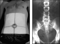
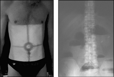
AP supine
• Patient lies supine upon the X-ray table
• Median sagittal plane is perpendicular and the anterior superior iliac spines (ASIS) are equidistant to the table top
• A pad may be placed under the knees for support
• Ensure patients hands are by their side and not under the pelvis


