Imaging modality
Indications and advantages
Limitations
Abdominal radiographs (KUB, obstructive series)
To demonstrate free air, bowel obstruction, calculi/calcification, foreign body, soft tissue mass displacing the bowel loops, ancillary findings like bone involvement
– Has a low sensitivity and specificity
– Poor anatomic localization
Fluoroscopy
To demonstrate bowel motility, strictures, gastric reflux, gastric emptying
In post op cases to demonstrate anastomotic leaks
US
– Noninvasive and portable
– Subjective and operator-dependent
– No radiation
– Decreased accuracy in patients with large body habitus
– First imaging modality for
– Low sensitivity and specificity
• Right upper quadrant
• Female pelvis
• Pediatric patients
CT
– High resolution
– Exposure to ionizing radiation
– Wide coverage
– Limited application in patients with contrast allergies or abnormal renal function
– Rapid investigation
– 24/7 Access
MRI
– Focused examination
– Limited availability
– Ideal for biliary/common bile duct evaluation
– Cost
– Imaging of choice for extended evaluation of pelvic pathology in women
– Claustrophobia
– Useful in pregnant patients
– Monitoring of young patients with Crohn’s disease to avoid excessive radiation
Imaging Techniques: Overview
Plain Radiography/Fluoroscopy
Imaging evaluation of patients presenting with clinical signs and symptoms localizing to the diseases of the abdomen often begins with an abdominal radiograph or kidney–ureter–bladder (KUB) radiograph or an obstructive series of radiographs (usually consisting of either erect and supine or lateral decubitus and supine abdomen images). Plain radiography is an inexpensive imaging technique that is performed in patients with findings of an acute abdomen for the diagnosis of bowel obstruction, bowel perforation, or urinary stones. Plain radiographs also contribute to the routine care of patients to confirm the position of various tubes (nasogastric tube or drainage catheters) and as follow-up examinations in patients with ileus. Plain film radiography can be done rapidly and portably, allowing detection of several acute abdominal emergencies such as perforated viscus or bowel obstruction. Its disadvantages include lack of contrast resolution and an inability to differentiate various intra-abdominal structures.
Fluoroscopic imaging techniques performed after instillation of oral contrast agents is a great tool to evaluate diseases of both the GI and GU tracts. It is a dynamic imaging technique that is particularly valuable in detection of abnormalities affecting bowel motility. This includes evaluation of esophageal motility disorders in patients with dysphagia, heart burn, or chest pain. Fluoroscopic imaging with oral contrast is especially valuable in postoperative patients to assess the integrity of the gastrointestinal tract after bowel anastomotic surgery. Early identification of anastomotic leaks in the postoperative period allows a surgeon to plan appropriate early interventions. Conventional barium studies also can be used as first-line noninvasive diagnostic studies in patients with dyspepsia, weight loss, an abdominal mass, and partial obstruction. Double contrast techniques can provide good detail of the bowel mucosa allowing identification of ulcerations, small protrusions, and any strictures.
Ultrasound
Ultrasound (US) is a valuable noninvasive imaging technique that uses sound waves to create images of internal structures. This imaging technique does not expose the patient to ionizing radiation and is performed by placing an US probe (a transducer) onto the skin over the organ of interest. US provides real time gray scale images of various structures within the abdomen, including deeper organs. This imaging technique is routinely used to evaluate the gallbladder, liver, bile ducts, spleen, pancreas, kidneys, uterus, ovaries, and abdominal cavity.
The main advantages of US include:
1.
US is universally available, easy to use, and is easily performed portably at the patient’s bedside.
2.
The real time, dynamic nature of US allows evaluation of motion in real time, e.g., to monitor peristalsis, observe fetal movements in pregnant women, and examine the effects of maneuvers, e.g., Valsalva and Mueller maneuvers, and gravity on internal organs.
3.
The real time nature of US permits analysis of effects of graded compression on various structures to help determine tissue stiffness, i.e., rigid vs. soft. This use is not limited to the abdomen and in fact is a mainstay in evaluation of deep venous thrombosis in the limbs.
4.
It allows direct visualization of blood flow and pulsations within vessels and masses.
5.
US is an excellent tool to perform image guided interventional procedures.
US is suitable for both focused examination and for routine screening. As a screening tool, US is frequently used in the diagnosis of an obvious abdominal mass, peritoneal fluid collections such as ascites, and in the evaluation of hydrobilia or hydronephrosis. It is often performed as an initial imaging study for the evaluation of abdominal pain particularly in patients with right upper quadrant pain as it allows accurate diagnosis of acute cholecystitis. US plays an equally critical role in evaluation of lower abdominal pain and bleeding in women of childbearing age. In addition to allowing the detection of various conditions, US is a safe technique to guide aspiration and drainage of fluid collections/abscesses to determine their nature.
Recent advances in US include harmonic imaging, 3-dimensional (3-D) imaging, and sono-elastography [1, 2]. Harmonic imaging uses ultrasonic sound waves similar to US, but requires a broad transducer bandwidth. Harmonic imaging is superior to conventional US as it provides better lesion visibility, improves diagnostic confidence, and typically shows enhanced contrast by removing image noise. Harmonic imaging is generally useful for depicting cystic lesions, deep seated vascular structures, and those lesions containing echogenic tissues such as fat, calcium, or air.
3D volumetric sonography allows 3D reconstructions of anatomic structures from US images. It has shown great benefit in fetal ultrasonography because it permits easy identification of fetal anatomy and confident interpretation of congenital anomalies [1, 2]. 3D imaging is also routinely used to evaluate uterine abnormalities including depiction of congenital uterine anomalies and visualization of the position of intrauterine contraceptive devices. 3D imaging increases the confidence of needle placement during interventional procedures because it shows three planes simultaneously.
Despite these advantages, caution should be used when utilizing US for investigation. Obesity, the presence of dense overlying tissue and gaseous distention, can degrade US images because high frequency sound waves do not penetrate these tissues well. Poor penetration can limit the technical utility of abdominal ultrasound or even render it completely unable to visualize target organs. US is also operator-dependent, relying greatly on the skills and experience of the sonographer. Incorrectly performed scans can create a level of uncertainty in the interpretation of images and the diagnosis of pathology. Because of the comparatively narrow field of view produced by current transducers and the freehand method with which scans are performed, it is even possible to overlook pathology in some circumstances. Some have likened US to looking through a keyhole. Therefore it is of utmost importance to be sure the sonographer is well-trained and experienced. Of course, radiologists ultimately interpret the studies.
Multidetector CT
The advent of multidetector CT (MDCT) has provided impressive diagnostic benefits in the management of abdominal and pelvic disorders [3]. Utilization of MDCT scans in patients presenting with abdominal pain has undergone explosive growth. Today, over 20 % of patients presenting to an emergency department with abdominal complaints undergo a CT examination. This represents a nearly tenfold increase in CT utilization over a 12-year period [4]. The high contrast resolution of CT makes it the preferred modality for high accuracy evaluation of both solid and hollow visceral pathology in the abdomen and pelvis.
The main advantages of MDCT include:
1.
MDCT is universally available and easily accessible (24 hrs a day, 7 days a week).
2.
MDCT allows rapid image acquisition with an ability to scan multiple body parts at the same time.
3.
Standardized scanning protocols and image display patterns make interpretation easier.
4.
It can be used for screening as well as for focused problem solving.
5.
Its value can be augmented by administering oral and intravenous contrast, depending on the application.
6.
CT has the capability to obtain images rapidly in sequences through a volume of tissue. This capability allows CT to depict different phases of filling of the vascular system as in CT angiography.
Despite these benefits, one of the major drawbacks of CT imaging is exposure to ionizing radiation, raising concerns about its long-term mutagenic and carcinogenic effects particularly in young patients who undergo multiple CT examinations [5]. The radiation dose is often dependent on type of examination with increased exposure encountered in CT exams with multiple phases such as arterial, venous and delayed phase.
In order to answer many clinical questions, CT of the abdomen and pelvis requires injection of intravenous iodinated contrast material (IVCM). Contrast enhanced MDCT studies improve lesion detection and characterization and also allow differentiation of lymph nodes from vascular structures. On the other hand, non-contrast CT, i.e., without intravenous contrast, is often sufficient to diagnose urinary tract stones and to identify suspected intra-abdominal hematomas.
For most applications, oral contrast material (OCM) also is administered routinely prior to a CT of the abdomen and pelvis. Intraluminal oral contrast is essential to help differentiate abnormal mesenteric lymph nodes from unopacified bowel loops, to facilitate detection of bowel abnormalities, to identify bowel perforations or anastomotic leaks, and to distinguish bowel from some extra intestinal pathology such as abscesses.
The OCM chosen for a given application can be positive or negative/neutral as indicated by its density measurements on the scan. Positive OCM includes dilute liquid barium suspensions and water-soluble iodine solutions, e.g., Gastrograffin, both of which appear denser than water on the scan. Negative/neutral OCM includes water and low-density barium solutions such as Volumen. Positive OCM is adequate for most indications in the abdomen, but neutral OCM is preferred for dedicated examinations that target the bowel itself, i.e., CT enterography (CTE). Iodinated contrast also can be injected through a catheter into the urinary bladder to diagnose urinary bladder rupture in patients with pelvic trauma, i.e., CT cystography (Table 5.2).
Table 5.2
MDCT protocols for imaging of the abdomen and pelvis
MDCT protocols | Clinical indications | Additional points |
|---|---|---|
No oral contrast | High grade small bowel obstruction, unstable patients, CT angiographic studies, and suspected gastrointestinal bleed | Oral contrast can interfere with 3D imaging |
Rectal contrast | Appendicitis, diverticulitis | |
Intravenous contrast agent | Used in all indications except in most cases of ureteral stones/flank pain | Intravenous contrast agent opacifies the abdominal vasculature and provides useful information regarding the enhancement patterns of parenchymal organs and intestine |
Intravenous contrast can be given in doubtful cases of ureteral stones | ||
Faster injection rate of intravenous contrast | GI bleed/bowel ischemia/hemorrhage/vascular complications | |
Dual phase (arterial and venous) | Liver or pancreatic mass | |
Delayed phase | Hematuria and pelvic pathology |
Magnetic Resonance Imaging
Typically, MRI of the abdomen and pelvis is reserved for problem solving, usually as a targeted study with a specific question in mind. It is also the investigation of choice in patients with a history of allergy to iodinated contrast who, as a consequence, cannot undergo a contrast enhanced MDCT. MRI does have drawbacks since it is expensive relative to other diagnostic imaging examinations, and even with today’s wider and shorter bore scanners, claustrophobic patients may be unable to tolerate being in the bore of the scanner. In addition, the long scanning times required to complete a study make MRI undesirable for acutely ill patients and for patients who are unable to lie still during image acquisition.
The superior contrast and soft tissue resolution of MRI makes it an invaluable technique in visualization of the hepato-pancreato-biliary pathology. The enhanced contrast resolution of MR coupled with its ability to depict tissues using multiple sequences to accentuate different tissue parameters have elevated MR as the preferred imaging modality for characterization of hepatic lesions. For example, MR is excellent at detecting early hepatocellular carcinomas in patients with chronic parenchymal liver disease such as that resulting from Hepatitis B or C infections.
Furthermore, the recent availability of specific hepatobiliary contrast agents has improved MR’s detection of hepatic metastases. MR plays a crucial role in monitoring the response of known hepatic malignancies to systemic chemotherapy, trans-arterial chemo embolization (TACE), and percutaneous ablative therapies, e.g., radiofrequency ablation (RFA). MR is considered very accurate in assessment of pancreatic lesions, particularly cystic lesions, and to evaluate the pancreatic duct. Magnetic resonance cholangiopancreatography (MRCP) provides two dimensional (2D) and 3D views of the biliary tree and pancreatic ducts and so in many cases has supplanted standard ERCP for noninvasive evaluation of the biliary tree.
MR enterography (MRE), also called MR enteroclysis, is a focused MR examination dedicated to evaluation of small bowel pathology. It has gained acceptance in the past decade as a replacement to MDCT because of concerns about high radiation exposure from repeated MDCT studies in patients with inflammatory bowel disease. MRE provides exquisite detail of small bowel pathology and is accurate in the identification of extra intestinal complications such as abscess and fistula.
MRI precisely depicts uterine and adnexal pathology and so is integral to staging of uterine and ovarian malignancies. In addition, because MRI does not employ ionizing radiation, it can be used repeatedly in young individuals and in pregnant women without fear of carcinogenesis. MR is performed routinely for local staging of patients with prostate and rectal cancer, both before surgery and afterwards to monitor therapeutic response.
Imaging for Abdominal pain
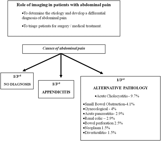
Abdominal pain is a common presentation and often the most challenging complaint to evaluate [5, 6]. This symptom accounts for nearly 2 % of outpatient visits and nearly 5 % of patients presenting to the emergency department. Most often the etiology of the pain is benign, but nearly 10 % of patients who present to the emergency department with abdominal pain have a life-threatening problem that often requires surgery (Fig. 5.1).
Evaluation of these patients requires a thorough and logical approach that depends on the location of the pain. For example, in conditions such as acute appendicitis or cholecystitis accurate pain localization has very strong predictive value [5]. Although imaging is often the final arbiter in the patient’s evaluation, it should not be considered as a substitute for a careful history and physical examination along with relevant laboratory tests (Tables 5.3 and 5.4). Imaging techniques such as plain radiographs or ultrasound should be considered as first-line tools before opting for advanced techniques such as CT or MRI [5, 6].
Table 5.3
Various causes and imaging investigation of choice in abdominal pain
Symptom | Causes | Imaging modality of choice | Second most preferred modality | Other imaging modalities |
|---|---|---|---|---|
Right upper quadrant | Biliary: cholecystitis, cholelithiasis, cholangitis | Ultrasound | MDCT | MRI—biliary anomalies |
HIDA—functional evaluation of hepatobiliary system | ||||
Colonic: colitis, diverticulitis | ||||
Hepatic: abscess, hepatitis, mass | ||||
Renal: nephrolithiasis, pyelonephritis | ||||
Epigastric pain | Biliary: cholecystitis, cholelithiasis, cholangitis | Ultrasound | MDCT | |
Gastric: esophagitis, gastritis, peptic ulcer | ||||
Pancreatic: mass, pancreatitis | ||||
Left upper quadrant | Gastric: esophagitis, gastritis, peptic ulcer | MDCT | Ultrasound | |
Pancreatic: mass, pancreatitis | ||||
Renal: nephrolithiasis, pyelonephritis | ||||
Right lower quadrant |


