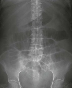
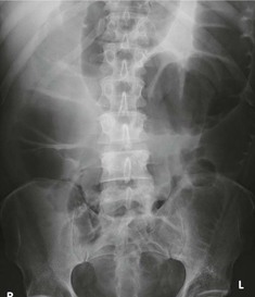
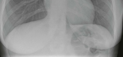
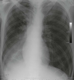
Abdominal Pain


Key Points


  • Point-of-care ultrasound assessment of unstable patients with abdominal pain should begin with an evaluation for hemorrhage, followed by an evaluation for infectious sources.


  • Point-of-care ultrasound assessment of stable patients with abdominal pain should focus on etiologies based on the patient’s complaints and demographics with an emphasis on time-sensitive emergencies.


  • Ultrasound is the initial diagnostic imaging modality of choice in children with abdominal pain because most etiologies can be safely and effectively diagnosed in this population without exposure to ionizing radiation.


Background


Point-of-care ultrasound has become an essential component in evaluation of patients with acute abdominal pain. Providers can integrate the clinical history and physical exam findings with a focused bedside ultrasound examination to guide diagnostic and therapeutic decisions. Point-of-care ultrasound results are immediately available, saving both time and personnel resources and avoiding the use of ionizing radiation or intravenous contrast agents.


A wide range of acute abdominal pathologies can be diagnosed with a focused abdominal ultrasound exam, including but not limited to the following: abdominal aortic aneurysm or dissection, appendicitis, renal or biliary colic, acute cholecystitis, small bowel obstruction, tubo-ovarian abscess, hemorrhagic or ruptured ovarian cyst, ascites, abdominal wall abscess, and bowel perforation ( Figure 39.1 ).


Figure 39.1


Point-of-care ultrasound is an indispensable tool for the evaluation of the patient with abdominal pain as it can facilitate the rapid diagnosis of a number of common emergent conditions. A, Abdominal aortic aneurysm. A 5-cm abdominal aortic aneurysm (AAA) containing mural thrombus (asterisk). B, Hemoperitoneum. Trace amount of free fluid (asterisk) in Morison’s pouch after liver laceration. C, Acute cholecystitis. Note the gallbladder (gb) with a thickened wall, pericholecystic fluid, and gallstone lodged in the gallbladder neck (asterisk). D, Acute appendicitis. The appendiceal tip is surrounded by hyperechoic peri-appendiceal fat stranding (asterisks). E, Obstructive uropathy. Anechoic signal within the renal pelvis (asterisks) consistent with mild to moderate hydronephrosis in a patient with an obstructing ureteral stone. F, Ruptured ectopic pregnancy (ep) with extrauterine gestation visualized deep to the uterus. There is associated complex free fluid (asterisks) representing hemoperitoneum.


Previous chapters have addressed image acquisition and interpretation techniques of focused abdominal ultrasound exams. This chapter presents a framework to conceptualize integration of point-of-care ultrasonography into initial evaluation of urgent or emergent abdominal complaints. Providers may perform a focused evaluation of one organ, a combination of abdominal organs, such as bladder and kidneys, or a combination of organs from different systems. We review the most important applications in evaluation of unstable and stable patients with abdominal pain.


Unstable Patients


Initial evaluation of unstable patients with abdominal pain should focus on the limited number of conditions for which point-of-care abdominal ultrasound results will dramatically change management decisions. Ultrasound approach in these patients can be divided into two components: a search for hemorrhage and a search for an infectious source ( Figure 39.2 ).


Figure 39.2


A proposed ultrasound approach to the unstable patient with abdominal pain: the provider begins with a search for hemorrhage (a Focused Assessment with Sonography in Trauma (FAST) exam and an abdominal aortic aneurysm evaluation) followed by a search for an infectious source of sepsis (acute cholecystitis and obstructive uropathy).


Hemorrhage


Evaluation of unstable patients with abdominal pain begins with a search for hemoperitoneum, best performed with the abdominal views of the Focused Assessment with Sonography in Trauma (FAST) exam. While most providers are aware that unstable trauma patients with positive FAST exams should undergo immediate operative exploration, hemoperitoneum can result from nontraumatic etiologies, including ruptured ectopic pregnancy or ovarian cysts, aneurysmal disease of the aorta or visceral vessels, anticoagulant use, blood dyscrasias, or tumor-associated hemorrhage. Detection of free fluid in Morison’s pouch, splenodiaphragmatic space, or the pelvis is critical to promptly identify patients in whom aggressive resuscitation with blood products is indicated while arranging definitive care ( Figure 39.3 ).


Figure 39.3


Approach to acute abdominal pain using point-of-care ultrasound in unstable and stable patients.


Evaluation for abdominal aortic aneurysm is included in the bedside abdominal ultrasound exam for hemorrhage. Given that the majority of ruptured abdominal aortic aneurysms (AAAs) cause retroperitoneal hemorrhage, the role of point-of-care ultrasound is more to detect AAAs that are large or dissecting rather than to detect intraperitoneal hemorrhage. Rapid identification of an AAA with suspected or impending rupture can dramatically expedite definitive management for patients that might otherwise have broad differential diagnoses. The abdominal aorta must be visualized from the diaphragm through its bifurcation into common iliac arteries in both long and short axes. While an abdominal aortic diameter >3 cm is considered aneurysmal, it is unusual for aneurysms <4.5 cm to rupture, and clinicians must consider the entire clinical presentation when incorporating ultrasound findings into decision making.


Patient demographics play an important role in evaluation of unstable patients. For example, a young female is far more likely to have a ruptured ectopic pregnancy than an abdominal aortic aneurysm, and clinicians should individualize their diagnostic approach accordingly.


Infection


Point-of-care abdominal ultrasound can play an important role in identifying an infectious source in unstable patients with septic shock. The gallbladder should be assessed for signs of acute cholecystitis, including wall thickening, pericholecystic fluid, stones, and/or a sonographic Murphy’s sign. Assessment of the common bile duct for dilation can confirm a suspicion of acute cholangitis. Unilateral hydronephrosis with or without a visualized ureteral stone can indicate obstructive uropathy with associated pyelonephritis. Intra-abdominal abscess, perforated appendicitis, small bowel obstruction, and pneumoperitoneum can be identified with point-of-care ultrasound and guide management, including prompt initiation of appropriate antibiotics and surgical consultation.

Only gold members can continue reading. Log In or Register to continue

Stay updated, free articles. Join our Telegram channel

May 20, 2019 | Posted by in ULTRASONOGRAPHY | Comments Off on Abdominal Pain

Full access? Get Clinical Tree

Get Clinical Tree app for offline access