Abdominal Wall Neoplasms
R. Brooke Jeffrey, MD
Key Facts
Terminology
Malignant abdominal wall tumor, not arising from normal constituents of abdominal wall
Imaging
Best diagnostic clue
Spherical soft tissue density mass(es) within abdominal wall muscles or subcutaneous tissues
Best imaging tool: CECT, US, or MR
Protocol advice
Always consider hernia before performing biopsy of abdominal wall mass
Top Differential Diagnoses
Abscess
Hematoma
Hernia
Varices
Endometriosis
Pathology
10% of patients with Gardner syndrome present with abdominal wall neoplasms
Clinical Issues
Most common signs/symptoms
Palpable mass
May be asymptomatic (lipoma or desmoid) or painful (metastases)
Clinical profile
Patient with known malignancy or lymphoma (especially immunocompromised patients)
Diagnostic Checklist
Consider spigelian hernia if mass is lateral to rectus sheath
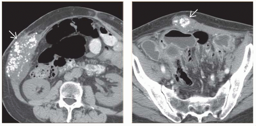 (Left) Axial NECT in a patient with a palpable right flank mass 2 years after a left colectomy for stage III mucinous adenocarcinoma of the descending colon. Note the densely calcified right flank mass  , proven on biopsy to be a metastasis of colon cancer. (Right) Axial NECT in the same patient demonstrates a calcified midline mass , proven on biopsy to be a metastasis of colon cancer. (Right) Axial NECT in the same patient demonstrates a calcified midline mass  . Given the fact that the lesion arose in close proximity to the prior surgical incision, this possibly represents a tumor implant at the time of colectomy. . Given the fact that the lesion arose in close proximity to the prior surgical incision, this possibly represents a tumor implant at the time of colectomy. |
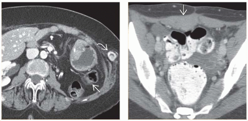 (Left) Axial CECT in a 50-year-old woman undergoing routine surveillance after a left nephrectomy for stage IIA renal cell carcinoma reveals bowel loops  occupying the left renal fossa. Note the hypervascular abdominal wall mass occupying the left renal fossa. Note the hypervascular abdominal wall mass  seen laterally, which was proven on biopsy to be a metastasis from renal cell carcinoma. (Right) Axial CECT in a 19-year-old patient with Gardner syndrome reveals a solid mass seen laterally, which was proven on biopsy to be a metastasis from renal cell carcinoma. (Right) Axial CECT in a 19-year-old patient with Gardner syndrome reveals a solid mass  involving the lower rectus muscle, surgically proven to be a desmoid tumor. involving the lower rectus muscle, surgically proven to be a desmoid tumor. |
TERMINOLOGY
Definitions
Benign or malignant abdominal wall tumor not arising from or metastatic to normal constituents of abdominal wall, including skin, subcutaneous fat, or muscle
IMAGING
General Features
Best diagnostic clue
Spherical soft tissue density mass(es) within abdominal wall muscles or subcutaneous tissues
Size
Few mm to several cm
Morphology
Spherical or oblong
Imaging Recommendations
Best imaging tool
CECT, US, or MR
Protocol advice
Always consider hernia before performing biopsy of abdominal wall mass
Radiographic Findings
Radiography
Abdominal wall calcifications within metastases from adenocarcinoma and serous cystadenocarcinoma of ovary
CT Findings
Lipoma
Subcutaneous fatty mass
Most primary tumors and metastases are enhancing, well-defined soft tissue masses
Some sarcomas and metastases are hypervascular
Renal cell carcinoma
Both mucinous colon cancer and serous ovarian carcinomas may calcify
Dermoids
Soft tissue attenuation
Often infiltrative appearance
Related posts:

Stay updated, free articles. Join our Telegram channel

Full access? Get Clinical Tree


