KEY FACTS
Terminology
- •
Most common nonlethal skeletal dysplasia
Imaging
- •
Long bones
- ○
Short limbs with normal ossification, no fractures
- ○
Rhizomelia: Proximal long bones (femur, humerus) more affected than distal long bones
- ○
Upper extremities more severely affected than lower
- ○
Femurs may show mild bowing but no angulation
- ○
Shortening often not present until late 2nd and 3rd trimester
- ○
- •
Head and face
- ○
Progressive macrocephaly with frontal bossing
- ○
Depressed nasal bridge with upturned nasal tip
- ○
- •
Chest normal to mildly bell-shaped
- •
Spine has prominent thoracolumbar kyphosis
- •
Trident-shaped hands with short fingers
Clinical Issues
- •
Autosomal dominant inheritance
- ○
If one parent has achondroplasia, fetus has 50% chance of being affected
- ○
- •
Over 80% of cases new mutations, so neither parent may be affected
Scanning Tips
- •
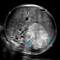
In any case of suspected skeletal dysplasia, pay particular attention to appearance and ossification of long bones
- •
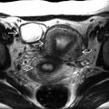
Profile views and 3D images of face are very important to look for classic features
- •
Obtain sagittal views of spine to look for abnormal outward curvature (kyphosis)
- •
Need to follow-up at-risk fetuses (i.e., parent with achondroplasia)
- ○
Limb shortening and classic facial features may not be apparent at time of routine anatomy scan
- ○
 and the depressed nasal bridge
  . The head circumference was > 95% for gestational age.
 .
.