KEY FACTS
Imaging
- •
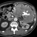
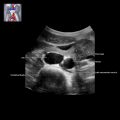
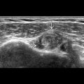
> 90% of fetal cases arise in adrenal gland
- •
May be solid, cystic, or mixed
- ○
Cystic appearance may represent involuting tumor and is excellent prognosis
- ○
- •
Calcification may be present but less common than in pediatric age group
- •
Liver most common location for metastases; may form discrete nodules or be diffusely infiltrating
- •
Look for placental metastases especially if maternal symptoms of preeclampsia
Top Differential Diagnoses
- •
Extralobar sequestration may present as suprarenal mass
- ○
Left-sided mass; separate normal adrenal gland
- ○
Dominant feeding vessel from aorta
- ○
- •
Adrenal hemorrhage much more common in pediatric population but has been reported in utero
- ○
No flow on Doppler; will involute over several weeks
- ○
Clinical Issues
- •
Variable fetal course
- ○
May resolve spontaneously, remain stable, or rarely progress to hydrops and even death
- ○
- •
Most fetal neuroblastomas have both favorable stage and biologic markers and have excellent prognosis
Scanning Tips
- •
Must perform careful, focused, high-frequency exam looking for normal adrenal gland
- ○
No normal adrenal gland supports neuroblastoma
- ○
If present, sequestration is more likely
- ○
- •
Assess vascularity with color Doppler
- ○
Some flow but no dominant feeding vessel
- ○
- •
Careful examination of liver for metastases
- ○
Diffusely infiltrating metastases are difficult to diagnose
- ○
Be suspicious when hepatomegaly or hydrops is present
- ○
- •
Close follow-up as may either grow or regress
- •
Monitor for hydrops
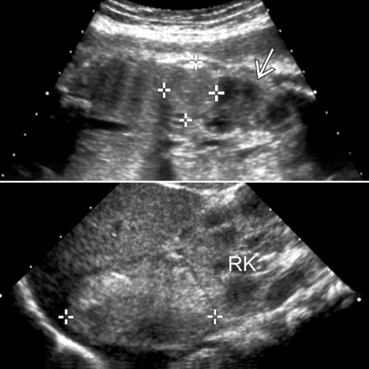
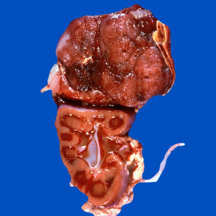
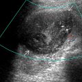
 . Sagittal ultrasound after delivery (bottom) confirms a solid, suprarenal mass (calipers).