GROSS ANATOMY
Overview
- •
Pancreas: Accessory digestive gland lying behind stomach in anterior pararenal space (APS) of retroperitoneum
- ○
Exocrine function: Pancreatic acinar cells secrete pancreatic juice → pancreatic duct → duodenum
- ○
Endocrine: Pancreatic islet cells (of Langerhans) secrete insulin, glucagon, and other polypeptides → portal venous system
- ○
Divisions
- •
Head: Thickest part; lies to right of superior mesenteric artery and vein (SMA, SMV)
- ○
Attached to “C” loop of duodenum (2nd and 3rd parts)
- ○
Uncinate process: Head extension, posterior to SMV
- ○
Bile duct lies along posterior surface of head, joins with pancreatic duct (of Wirsung) to form hepatopancreatic ampulla (of Vater)
- ○
Main pancreatic and bile ducts empty into major papilla in 2nd portion of duodenum
- ○
- •
Neck: Thinnest part; lies anterior to SMA, SMV
- ○
SMV joins splenic vein behind pancreatic neck to form portal vein
- ○
- •
Body: Main part; lies to left of SMA, SMV
- ○
Splenic vein lies in groove on posterior surface of body
- ○
Anterior surface is covered with peritoneum forming back surface of omental bursa (lesser sac)
- ○
- •
Tail: Lies between layers of splenorenal ligament in splenic hilum
Internal Structures
- •
Pancreatic duct (of Wirsung) runs length of pancreas, turning inferiorly through head to join bile duct
- •
Accessory pancreatic duct (of Santorini) opens into duodenum at minor duodenal papilla
- ○
Usually communicates with main pancreatic duct
- ○
Variations are common, including dominant accessory duct draining most pancreatic juice
- ○
- •
Vessels, nerves, and lymphatics
- ○
Arteries to head mainly from gastroduodenal artery
- –
Pancreaticoduodenal arcade of vessels around head also supplied by SMA branches
- –
- ○
Arteries to body and tail from splenic artery
- ○
Veins are tributaries of SMV and splenic vein → portal vein
- ○
Autonomic nerves from celiac and superior mesenteric plexus
- –
Parasympathetic stimulation of pancreatic secretion, but pancreatic juice secretion is mostly under hormonal control (secretin, from duodenum)
- –
- ○
Lymphatics follow blood vessels
- –
Collect in splenic, celiac, superior mesenteric and hepatic nodes
- –
- ○
IMAGING ANATOMY
Overview
- •
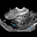
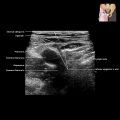
Pancreas can be localized on ultrasound by
- ○
Typical parenchymal architecture: Homogeneously isoechoic/hyperechoic echo pattern when compared with overlying liver
- ○
Surrounding anatomical landmarks: Body anterior to splenic vein; neck anterior to SMA/SMV
- ○
- •
Variations in reflectivity related to degree of fatty infiltration; uncinate process and posterior pancreatic head are relatively echo poor in 25% of subjects (lack of intraparenchymal fat)
ANATOMY IMAGING ISSUES
Imaging Recommendations
- •
Use 2- to 5-MHz transducers or up to 9 MHz for smaller patients
- •
Techniques to combat overlying stomach and bowel gas include
- ○
Displacement of intervening bowel gas by gentle graded compression with transducer
- ○
Overnight fasting or fasting > 6-8 hours
- ○
Noneffervescent fluid can be given orally to fill gastric fundus
- –
Scanning delayed for few minutes to allow fluid to settle
- –
Patient can lie on left side to allow imaging of body and tail of pancreas
- –
Patient can then be turned right to allow gastric fluid to flow to stomach antrum and duodenum, allowing imaging of head and uncinate process
- –
- ○
- •
CT is preferred imaging modality for imaging of pancreas
- •
MRCP (± secretin) or ERCP useful for defining pancreatic duct
Imaging Pitfalls
- •
Ultrasound examination of pancreas is often limited by overlying bowel gas
Key Concepts
- •
Shape, size, and texture of pancreas are quite variable
- ○
Largest in young adults
- ○
Atrophy and fatty infiltration with age (> 70), obesity, diabetes, corticosteroids, Cushing disease
- ○
Pancreatic duct also becomes more prominent with age (normal < 3 mm diameter)
- ○
Focal bulge or mass effect is abnormal
- ○
- •
Location behind lesser sac
- ○
Acute pancreatitis often results in lesser sac fluid (may mimic pseudocyst)
- ○
- •
Pancreas lies in APS
- ○
Inflammation (from pancreatitis) easily spreads to duodenum and descending colon, which are also located in APS
- ○
Inflammation easily spreads into mesentery and mesocolon; roots of these lie just ventral to pancreas
- ○
- •
Obstruction of pancreatic duct
- ○
Relatively common result of chronic pancreatitis (fibrosis &/or stone occluding pancreatic duct) or pancreatic ductal carcinoma
- ○
- •
Acute pancreatitis
- ○
Relatively common result of gallstone (lodged in hepatopancreatic ampulla causing bile to reflux into pancreas) or damage from alcohol abuse
- ○
PANCREAS IN SITU