TERMINOLOGY
Abbreviations
- •
Origin (O), insertion (I)
Definitions
- •
Junctional region between thigh and trunk
IMAGING ANATOMY
Overview
- •
Incorporates lower abdominal wall, inguinal canal, femoral triangle, and femoral adductor muscles
Osseous Anatomy
- •
Pubic bone
- ○
Pubic tubercle : Small protuberance, lateral border pubic crest
- –
Attachment: Inguinal ligament
- –
- ○
Superior and inferior rami : Extend from pubis
- ○
Pecten : Ridge, posterior aspect superior pubis
- –
O of pectineus muscle
- –
I of conjoint tendon (internal oblique and transverse abdominis)
- –
- ○
Pubic crest : Superior surface, anterior aspect pubic body
- –
O of rectus abdominis muscle
- –
I of transversus abdominis and external oblique muscles
- –
- ○
Pubic symphysis : Cartilaginous joint between pubic bodies
- ○
Superior pubic ligament : Laterally extends to pubic tubercles
- ○
Anterior Abdominal Wall
- •
Abdominal wall muscles
- ○
Rectus abdominis : Paired midline muscles
- –
O: Superior pubic ramus, pubic crest
- –
I: Xiphoid process, costal cartilages 5-7
- –
- ○
Linea alba : Aponeurotic junction of rectus femoris, transverse abdominis, and internal and external oblique muscles
- ○
External oblique : Most superficial
- –
O: Ribs 5-12
- –
I: Pubic crest, anterior iliac crest, linea alba
- –
Lower border of aponeurosis contributes to inguinal ligament
- –
- ○
Internal oblique : Between external oblique, transversus abdominis
- –
O: Lateral inguinal ligament, iliac crest, thoracolumbar fascia
- –
I: Pecten (conjoined tendon), pubic crest, inferior aspect of ribs 10-12, linea alba
- –
O: Anterior to deep inguinal ring
- –
I: Lateral to rectus abdominis muscle, posterior and medial to superficial inguinal ring
- –
Arches over inguinal canal, forming roof
- –
- ○
Transversus abdominis : Deepest
- –
O: Iliac crest, posterior aspect of lateral inguinal ligament, thoracolumbar fascia
- –
I: Pubic crest, pecten (conjoined tendon), linea alba
- –
Remains posterior to inguinal canal
- –
- ○
Inguinal Ligament
- •
Thickening inferior border of external oblique aponeurosis
- •
Attachments: Anterior superior iliac spine and pubic tubercle
- •
Separates lower extremity from pelvis
- •
Fascia lata attaches to inferior border
- •
Subinguinal space : Deep to inguinal ligament
- ○
Passageway for femoral vessels and nerve, iliopsoas muscle into femoral triangle
- ○
External iliac vessels become femoral vessels upon entering this space
- ○
Inguinal Canal
- •
Entrance: Deep inguinal ring
- ○
Located midinguinal ligament
- ○
Opening of evaginated transversalis fascia through which spermatic cord/round ligament pass
- ○
- •
Exit: Superficial inguinal ring
- ○
Division of external oblique aponeurosis lateral to pubic tubercle
- ○
- •
Contents: Ilioinguinal nerve; small arteries and veins; male: Spermatic cord; female: Round ligament
- ○
Covered by evaginated transversalis fascia
- ○
Adductor Musculature
- •
Adductor longus muscle : Thin tendon arises from medial superior pubic ramus
- ○
Overlies O of gracilis, adductor brevis, and magnus muscles
- ○
- •
Gracilis muscle : O from anterior aspect of symphysis pubis and medial aspect of inferior pubic ramus
- ○
O medial to adductor brevis muscle, deep to adductor longus muscle
- ○
Groin Lymph Nodes
- •
Superficial inguinal nodes
- ○
Lie in subcutaneous tissues below inguinal ligament and alongside great saphenous vein
- ○
Subdivided into 3 groups of lymph nodes
- –
Inferior group are located distal to saphenous opening and receive drainage from lower limb
- –
Superolateral group are located lateral to saphenous opening and receive drainage from lateral buttock and lower anterior abdominal wall regions
- –
Superomedial group are located medial to saphenous opening and receive drainage from perineum and external genitalia
- –
- ○
- •
Deep inguinal nodes
- ○
3-5 nodes located in femoral triangle just medial to femoral vein
- ○
Cloquet node is most proximal of deep inguinal lymph nodes located just below inguinal ligament
- ○
ANATOMY IMAGING ISSUES
Imaging Recommendations
- •
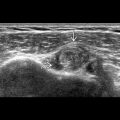
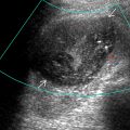
Usual imaging pathway for assessing groin pathology includes radiography &/or ultrasound
- •
Use curvilinear abdominal transducer for deeper structures in addition to/or high-resolution linear transducers
- •
Dynamic imaging is important, particularly in assessment of hernias or other position dependent pathology
GROIN