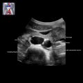
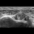
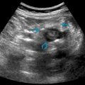
KEY FACTS
Terminology
- •
Benign, fluid-filled nonneoplastic renal lesion not meeting criteria of simple renal cyst, Bosniak classes II, IIF, and III
Imaging
- •
Round, oval, or irregular-shaped anechoic lesion
- •
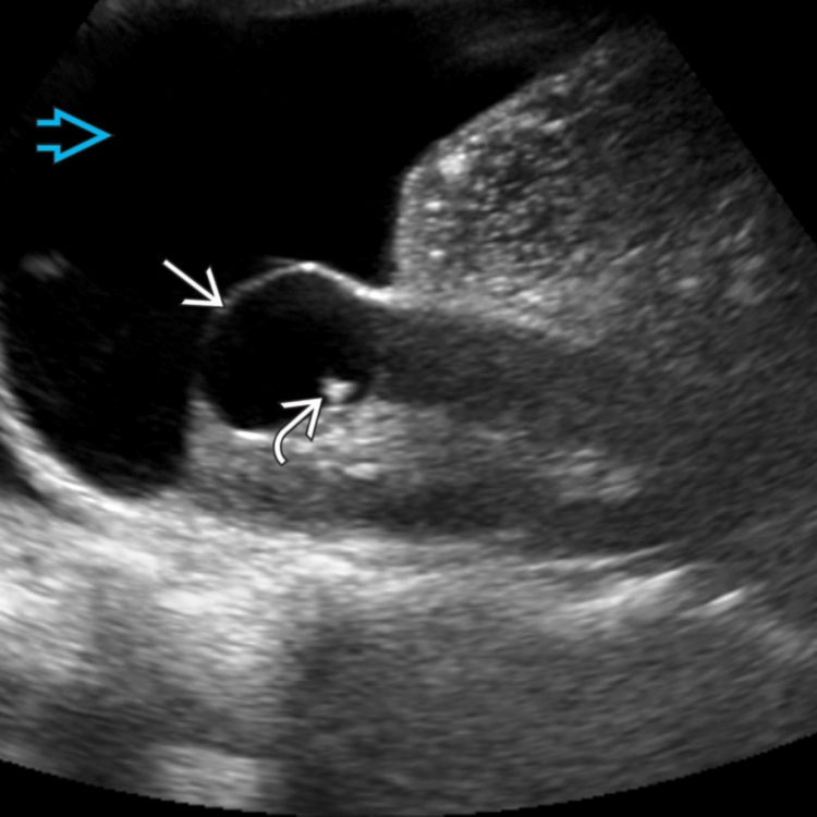
Hemorrhagic cyst: Variable → echogenic fluid, retracting clot, debris level or septations
- •
Proteinaceous cyst: Low-level echoes with bright reflectors or even layers of echoes
- •
Infected cyst: Thick wall with scattered internal echoes ± debris-fluid level
- •
Calcified cyst: Wall or septal calcification ± shadowing
- •
Neoplastic features: Solid mural or septal nodules, irregular wall, or irregular septal thickening with color flow
- •
Complex cysts should be evaluated with CEUS, CECT, or CEMR for decision of surgical intervention
- •
Contrast uptake on CEUS suspicious for malignancy (other than a few bubbles in thin, smooth septa or wall)
- ○
Increased sensitivity for detecting malignancy
- ○
- •
Information analogous to Bosniak classification
- •
Depending on body habitus and number of cysts, US can fully characterize renal cysts/monitor complex renal cysts
Top Differential Diagnoses
- •
Renal cell carcinoma (cystic)
- •
Multilocular cystic nephroma
- •
Localized cystic disease
- •
Renal abscess
- •
Renal metastasis/lymphoma
- •
Renal lymphoma
Clinical Issues
- •
20-30% of middle-aged adults; > 50% of patients > 50 years
- •
Complications: Hydronephrosis, hemorrhage, infection, cyst rupture
Scanning Tips
- •
Optimize color Doppler settings to detect flow in septa and nodules
 with a thin, smooth septation
  . Posterior acoustic enhancement is present
  .
.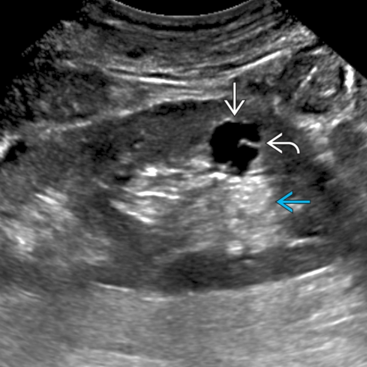
 of this minimally complex cyst.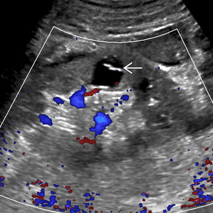
 , which did not have flow on color Doppler. There are no internal nodules, wall, or septal thickening; this corresponds to a Bosniak II lesion.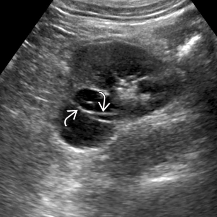
 . After injecting contrast, there was no internal flow
  , confirming that this was an intracystic hemorrhage. The other cysts
  were simple.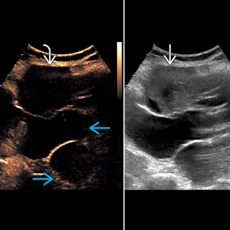
 . However, there was no color flow in the solid component. Multiple other cysts
  are present.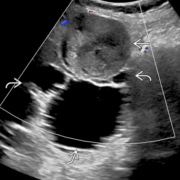
 with a small echogenic peripheral nodule
  . Color Doppler (not shown) did not show internal flow. There is incidentally a large amount of ascites
  .
.