KEY FACTS
Terminology
- •
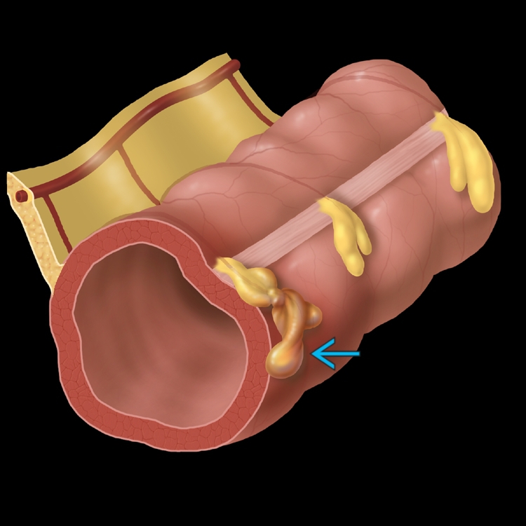
Ischemic infarction of epiploic appendages (small pouches filled with fat, located along colon)
Imaging
- •
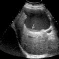
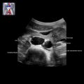
Noncompressible, hyperechoic oval mass, adjacent to colon, deep to region of maximal tenderness
- •
Adjacent absent or minimal bowel wall thickening with local mass effect
- •
Hypoechoic rim of inflamed visceral peritoneum (93%)
- •
± central hypoechoic areas of hemorrhagic change
- •
Color Doppler: Absence of central blood flow
- •
Contrast-enhanced ultrasound
- ○
Rim of peripheral arterial hyperenhancement
- ○
Central, nonenhancing hypoechoic regions
- ○
Top Differential Diagnoses
- •
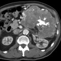
Segmental omental infarction
- •
Diverticulitis
- •
Appendicitis
- •
Sclerosing mesenteritis
- •
Primary tumors and mesocolon metastases
- •
Pelvic inflammatory disease
Pathology
- •
Torsion of epiploic appendage along its long axis with impairment of its vascular supply and subsequent necrosis
- •
Rectosigmoid junction is most common site
Clinical Issues
- •
4th-5th decades of life; male predominance (M:F = 4:1)
- •
Abrupt onset of very localized abdominal pain, most frequently left lower quadrant, gradually resolving over 3-10 days, palpable mass (10-30%)
- •
Mild or absent systemic symptoms and signs
- •
Obesity and strenuous exercise are recognized risk factors
- •
Conservatively managed
- •
Usually self-limiting condition with clinical recovery within 10 days
Scanning Tips
- •
Use combination of linear and curved transducers over area of maximal pain and tenderness
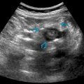
 and 2 adjacent normal appendages.