EMBRYOLOGY AND ANATOMY
Overview
- •
Trachea separates from foregut and developing esophagus
- ○
Failure to separate results in tracheoesophageal fistula
- ○
- •
Lungs go through several stages of development from primitive bronchi to alveoli capable of respiration
- ○
Until alveoli develop (~ 25 weeks gestational age) fetus would not be able to breath if delivered
- ○
Adequate intrathoracic volume required for normal pulmonary development
- –
Intrathoracic masses (especially diaphragmatic hernia) and chest wall abnormalities (e.g., skeletal dysplasias) restrict space for lung growth
- –
- ○
Amniotic fluid and fetal breathing also required for normal lung development; oligohydramnios has severe adverse effect on lung development
- ○
- •
Diaphragm development is complex with 4 embryologic structures which must fuse; failure of fusion results in a congenital diaphragmatic hernia (CDH)
SCANNING APPROACH AND IMAGING ISSUES
Techniques and Sonographic Appearance
- •
Required chest views include: 4-chamber heart, outflow tracts, and diaphragm
- ○
Evaluation of lungs is not specified but are generally well seen when obtaining required views
- ○
- •
Heart occupies ~ 1/3 of intrathoracic area
- ○
Cardiothoracic ratio is calculated by dividing cardiac circumference by thoracic circumference (TC); normal being ~ 50%
- ○
Increased ratio usually indicates cardiomegaly but may also occur when chest is small
- ○
- •
Lungs are initially similar to liver in echogenicity but become more echogenic with advancing gestation
- ○
Any aberration in this homogeneous echotexture raises suspicion for mass
- ○
- •
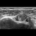
Diaphragm appears as a thin, arched, hypoechoic band
- ○
Best imaged in sagittal plane; need to assess both sides completely moving from chest wall to chest wall during real-time scanning
- –
Most CDHs occur posteriorly; if only viewed in anterior coronal plane, a CDH may be missed
- –
- ○
- •
Thymus may be prominent in 3rd trimester
- ○
Above the heart and is hypoechoic compared to lungs with a slightly reticular appearance
- ○
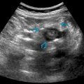
Look for thy-box to confirm it is not a chest mass
- –
Color Doppler of superior mediastinum, at level of 3-vessel view, shows thymus between internal mammary arteries creating box appearance
- –
- ○
- •
Fetal breathing is essential for normal lung development and is one component of biophysical profile
Approach to Fetal Chest Mass
- •
The following questions form a framework for evaluation of the fetal chest; specific diagnoses will be discussed in other chapters
- •
Is the chest normal in size?
- ○
A TC is not generally performed unless there is concern that the chest is small (e.g., skeletal dysplasia)
- –
Performed at level of 4-chamber view with soft tissues excluded
- –
- ○
Can compare to expected value for gestational age or as a ratio with the abdominal circumference (AC)
- –
TC:AC ratio is stable throughout gestation with normal being > 0.8
- –
Enlarged chest size is unusual but is a prominent feature of congenital high airway obstruction sequence (CHAOS)
- –
- ○
- •
Is the axis of the heart deviated?
- ○
Any shift in cardiac axis is highly suspicious for a thoracic mass or, alternatively, a cardiac defect
- ○
While a normal axis rules out most significant chest masses, small masses may not deviate axis
- ○
- •
Where is the stomach?
- ○
Absence of the normal abdominal stomach bubble is a cardinal sign of a left-sided CDH (stomach in chest)
- ○
Important to note, however, that a left-sided hernia may contain only bowel &/or liver, with stomach remaining below diaphragm
- ○
With a right-sided CDH, stomach remains in the abdomen but is often more midline than normal
- ○
- •
Is the mass cystic or solid?
- ○
Although there is overlap in the 2 differentials [congenital pulmonary airway malformation (CPAM) and CDH may look either cystic or solid], this is the starting point for forming a differential diagnosis
- ○
- •
If cystic, is it a simple cyst or a complex cystic mass?
- ○
Simple cyst is more likely to be a foregut duplication cyst, while a complex cystic mass is more likely to be a CDH, macrocystic CPAM, or lymphangioma
- ○
Do not confuse an effusion with a cystic mass; lung will float within an effusion and have a wing-like appearance, while a cystic mass will displace and compress lung
- ○
- •
If solid, what does the Doppler show?
- ○
CPAM: Vascular supply from pulmonary circulation
- ○
Sequestration: Prominent feeding vessel from aorta
- ○
CDH containing liver will show hepatic and portal veins
- ○
- •
Where is the mass?
- ○
Sequestrations are almost always at left lung base (or below the diaphragm), while CPAMs are more variable in location, occurring equally on both sides
- ○
Bilateral chest masses are less common but include bilateral CPAMs, bilateral CDHs, or CHAOS
- ○
- •
Does the mass extend beyond the chest wall?
- ○
Lymphangiomas are primarily in subcutaneous tissues, with secondary intrathoracic involvement
- ○
Teratomas can be locally aggressive and erode through chest wall
- ○
- •
Is there hydrops?
- ○
Development of hydrops is a poor prognostic sign and may warrant clinical intervention (e.g., cyst drainage, in utero resection, early delivery); all chest masses should be monitored carefully for developing hydrops
- ○
- •
Are there other anomalies?
- ○
CDH has a high association with chromosomal anomalies, other structural anomalies, and syndromes
- ○
Especially important to carefully evaluate the heart
- ○
Because the cardiac axis is often distorted, it may be more difficult to adequately evaluate, and a dedicated fetal echo may be warranted
- ○
LUNGS, DIAPHRAGM, & THYMUS