Acute Abdomen and Abdominal Tuberculosis
USG is advantageous because of its dynamic and real-time nature.
Can observe the presence or absence of peristaltic movements, blood flow pulsations, and fetal movements. Effects of valsalva, compression, and so on can be easily visualized.
High-frequency probe delineates the abdomen better. An expeditious evaluation—considering patient’s age, gender, associated symptoms, duration, and location of pain—is imperative to rule out crucial pathologies requiring emergent management.
Most of the etiologies are discussed in the respective sections.
Etiologies:
1. Appendicitis.
2. Perforated peptic ulcer: Free fluid can be seen.
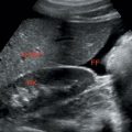
3. Pelvic inflammatory disease (PID): Free air can be seen to collect between the liver and the lateral abdomen wall.
4. Adnexal lesion.
5. Ectopic pregnancy.
6. Ruptured aortic aneurysm.
7. Ureteral stone.
8. Renal colic.
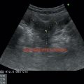
9. Crohns disease: Marked thickening of bowel wall in a skip pattern.
May be associated with focal discontinuity of the wall and a small walled-off abscess.
Surrounding inflammation of fat, seen as hyperechoic noncompressible tissue adjacent to cecum. Mesenteric lymphadenopathy may be associated.
10. Infections ileocolitis/terminal ileitis.
S/s—diarrhea, abdominal pain.
Diffuse thickening of terminal ileum and cecum.
Appendix appears normal.
Mesenteric lymphadenopathy.
11. Mesenteric lymphadenitis:
Stay updated, free articles. Join our Telegram channel

Full access? Get Clinical Tree


