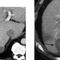
4 Acute Abdominal Trauma The most common cause of morbidity and mortality in the second, third, and fourth decades of life is trauma. The leading cause is automobile accidents, with penetrating injury, battery, and sports-related injury also important factors. The prevalence of trauma and cost to society are increasing. The spectrum of major trauma encompasses neurological, thoracic, abdominal, and musculoskeletal injuries. Life-threatening consequences may result from injury to any body region. Blunt abdominal injury poses a particular problem because there may be minimal physical findings, particularly if the patient’s state of consciousness is impaired. In addition, blunt abdominal injury may coexist with other significant trauma, including intracranial hemorrhage, ruptured thoracic aorta and pneumothorax, and musculoskeletal injury (e.g., major spinal, pelvic, and long-bone fractures). An accurate evaluation of abdominal injury is required to select the patient’s management. With the advent of sequential computed tomographic (CT) scanning for blunt abdominal injury in the late 1970s and early 1980s, surgeons began to refine their indications for exploratory laparotomy based on an evolving understanding of the natural history of blunt abdominal injury.1,2 In a hemodynamically stable patient, both major and minor lacerations of the liver, spleen, and kidneys will usually resolve with conservative management and no sequelae. Hepatic and splenic lacerations with intraperitoneal blood are not primary indications for laparotomy. Intervention, either angiographic or surgical, is usually reserved for lacerations associated with active bleeding, manifest as vascular contrast extravasation at the time of emergency CT scanning.3 Other CT findings in a hemodynamically stable patient that may prompt immediate laparotomy include intestinal rupture, central renal pedicle injury, pancreatic transection, and intraperitoneal bladder rupture. The major indication for laparotomy in a hemodynamically unstable patient is hemoperitoneum (without obvious extraabdominal blood loss) and hypotension unresponsive to intravenous fluid resuscitation. In the past, clinical evaluation and diagnostic peritoneal lavage determined the need for laparotomy.4 Sonography has largely supplanted diagnostic peritoneal lavage for detecting hemoperitoneum.5–8 CT is the preferred technique for evaluating normotensive patients with documented abdominal injury.9 It provides accurate global diagnostic information about abdominal and pelvic injury. However, the relatively high rate of negative abdominal and pelvic CT scans following blunt trauma, particularly in patients who are neurologically impaired, has prompted the use of sonography as a more cost-effective and efficient screening study.5–8 Hemo-dynamically stable patients with documented intraperitoneal fluid on sonography should proceed to CT evaluation. An abdominal sonogram that is negative for intraperitoneal fluid has been assumed to be sufficient to exclude significant blunt abdominal trauma in a patient without hematuria and with no clinical evidence of occult injury following 6 hours of observation.10 This chapter discusses the role of abdominal sonography and CT scanning in evaluating patients with suspected blunt abdominal injury. Both hemodynamically unstable and hemodynamically stable patients are discussed. The goal of sonographic imaging in trauma is to provide a cost-effective triage with high sensitivity and negative predictive value that leads to appropriate patient management and outcome. Intraperitoneal fluid may be blood, bile, urine, pancreatic juice, lymph, intestinal content, or preexistent ascites. Intraperitoneal fluid following blunt trauma is most often intraperitoneal blood resulting from hepatic, splenic, or mesenteric laceration. A periportal liver laceration or gall-bladder rupture may cause leakage of bile into the peritoneal cavity. In acutely injured patients, bile is usually not the predominant component of intraperitoneal fluid. Other types of intraperitoneal fluid in the patient with blunt trauma are unusual. Urinary ascites usually results from intraperitoneal rupture of the bladder. The dome of the bladder is the most common site of rupture. Pancreatic ascites may result when there is pancreatic transection associated with disruption of the posterior parietal peritoneum. Pancreatic ascites is usually caused by delayed intraperitoneal rupture of a pancreatic pseudocyst. Chylous ascites is very uncommon. It results from transection of the cisterna chyli or proximal thoracic duct. Patients with underlying liver disease with portal hypertension and hypoproteinemia may have preexisting ascites. Injuries to the liver and spleen result from compression or shearing force.11 The resultant lacerations may be localized or multifocal and may or may not be associated with a capsular tear. Grading schemes, such as CT injury severity scores, have been used to classify the extent of solid organ injury.12 However, there is no direct correlation between the injury severity score and the likelihood of delayed bleeding.12 Parenchymal and capsular tears usually result in a localized perihepatic or perisplenic hematoma, such as a sentinel clot,13 as well as intraperitoneal bleeding in the perihepatic or perisplenic spaces, paracolic gutters, or pelvis. This common pattern of hemoperitoneum distribution means that a rapid four-quadrant sonographic assessment will usually detect intraperitoneal fluid associated with hepatic or splenic injury. Injuries to the mesentery or bowel are more problematic. Hematoma may be confined to the bowel wall or mesentery without lateral or inferior distribution into the dependent intraperitoneal spaces.14 Because there may be no hemoperitoneum, an emergency sonogram may not detect these injuries. Extraperitoneal organ injury does not usually result in intraperitoneal bleeding. Injuries to the kidneys, pancreas, and duodenum, and disruption of the aortoiliac arterial system (caused by seat belt injury or pelvic fracture) are in this category. Thus, a negative sonogram for intraperitoneal fluid in patients with a central abdominal compression injury or hematuria does not exclude the possibility of significant injury.15 Patients with hematuria and those with lumbar spine or pelvic fractures should have an abdominal pelvic CT scan to exclude injuries even when sonography is negative.15 In addition, care must be taken to avoid missing traumatic pneumothorax, which has a relatively high association with blunt abdominal injury.16 Patients with abdominal injury are assessed for hemodynamic stability and the presence of associated injuries, including neurological, thoracic, and orthopedic injuries. Patients should be triaged into a hemodynamically unstable or hemodynamically stable category to guide further evaluation. If patients are unresponsive, either due to neurological injury or alcoholism, then external signs of abdominal trauma, including body wall contusion, should be assessed on a clinical examination. For patients who are conscious and responsive, clinical evaluation for focal guarding and rebound tenderness is vital. Plasma expanders are usually infused via intravenous lines. Bladder catheterization is used to evaluate for hematuria and to measure urine output. Tube thoracostomy is performed if any significant hemopneumothorax is present. Endotracheal intubation may be required for neurologically impaired patients or those in respiratory difficulty following aspiration or tube thoracostomy. The early triage diagnostic evaluation by sonography is usually performed concurrent with these diagnostic or resuscitative procedures. The hemodynamically unstable patient with suspected abdominal injury requires urgent evaluation for possible hemoperitoneum. Diagnostic peritoneal lavage can fulfill this role with very high reported sensitivity and specificity.4 Limitations of diagnostic peritoneal lavage include its inability to detect contained intracapsular hemorrhage in the liver or spleen, and relative insensitivity to bowel perforation and mesenteric hemorrhage. Diagnostic peritoneal lavage is insensitive to retroperitoneal injury, including pancreatic, duodenal, and renal injuries. A false-positive diagnostic peritoneal lavage may result from a traumatic peritoneal tap. Sonography can be performed as an alternative test to diagnostic peritoneal lavage.5,8 In general, sonography and diagnostic peritoneal lavage share the same limitations: suboptimal detection of solid organ injury, mesenteric hemorrhage, bowel perforation, and retroperitoneal injury.17 A positive diagnostic peritoneal lavage or positive sono-gram in the hemodynamically unstable patient should result in urgent laparotomy. It may be appropriate to delay laparotomy in some patients with multiorgan injuries. Severe associated neurological injury may require early evaluation by neurological CT. Thoracic injury may require portable chest radiography, tube thoracostomy, or a vascular phase multidetector CT examination to evaluate for possible thoracic aortic rupture. A negative diagnostic peritoneal lavage or negative sonogram in a hemodynamically unstable patient should lead to urgent evaluation of the thorax, abdominal retroperitoneum, pelvis, or extremities as sources of continuing blood loss. The hemodynamically stable patient may be assessed by sonography. Detection of intraperitoneal fluid or sentinel clot should be an indication for an abdominal pelvic CT study. A negative emergency abdominal sonogram, in the hemodynamically stable patient, does not exclude the possibility of contained intrahepatic or intrasplenic hematoma, mesenteric or bowel wall injury, or retroperitoneal injury.15
Differential Diagnosis
Intraperitoneal Fluid
Hepatic and Splenic Injury
Mesenteric and Bowel Injury
Extraperitoneal Organ Injury
Diagnostic Evaluation
Nonimaging Evaluation
The Hemodynamically Unstable Patient
The Hemodynamically Stable Patient
![]()
Stay updated, free articles. Join our Telegram channel

Full access? Get Clinical Tree


