Acute Aortic Intramural Hematoma
Katherine R. Birchard
CLINICAL HISTORY
69-year-old female with chest pain.
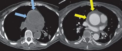 FIGURE 34A |
FINDINGS
Figure 34A: Axial noncontrast CT image shows near-circumferential high attenuation soft tissue (blue arrows) in the wall of a dilated ascending aorta; note that the blood in the lumen of the aorta is lower in attenuation than in the wall. Axial contrast-enhanced CT image at the same level shows contrast in the aortic lumen, and persistence of the abnormal soft tissue in the aortic wall (yellow arrows).
DIFFERENTIAL DIAGNOSIS
Acute intramural hematoma, atheromatous plaque, penetrating ulcer, chronic thrombus.
DIAGNOSIS
Acute aortic intramural hematoma.
Related posts:


Stay updated, free articles. Join our Telegram channel

Full access? Get Clinical Tree


