Acute Cholecystitis
Parth C. Patel
Ellie R. Lee
CLINICAL HISTORY
40-year-old female who presents with acute right upper quadrant abdominal pain, nausea, and vomiting.
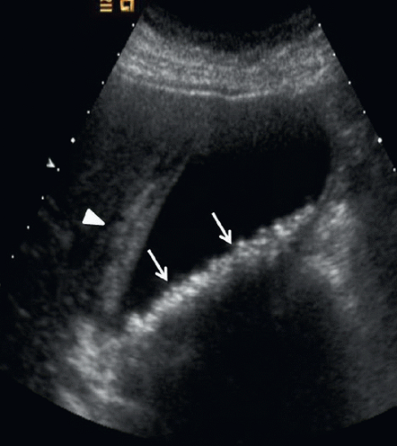 FIGURE 99A |
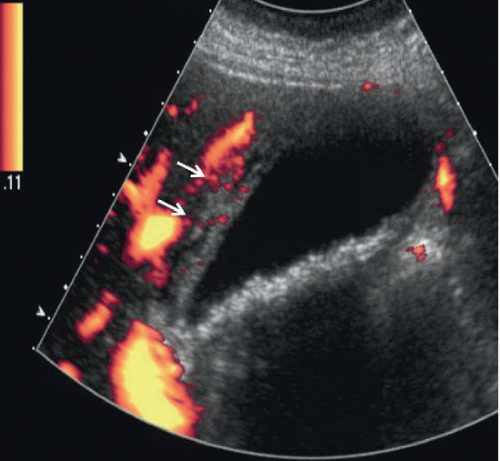 FIGURE 99B |
FINDINGS
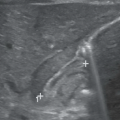
Figure 99A: Longitudinal ultrasound image of the gallbladder demonstrates multiple small shadowing gallstones (arrows) and thickened gallbladder wall measuring up to 1 cm (arrowhead). The sonographic Murphy’s sign was positive. Figure 99B: Longitudinal power Doppler ultrasound image of the gallbladder shows thickened, hyperemic gallbladder wall (arrows). Findings compatible with acute cholecystitis.
DIFFERENTIAL DIAGNOSIS
Chronic cholecystitis, acute hepatitis, hypoproteinemia, adenomyomatosis, pancreatitis.
DIAGNOSIS
Acute calculous cholecystitis.
DISCUSSION
Related posts:


Stay updated, free articles. Join our Telegram channel

Full access? Get Clinical Tree


