Pneumoperitoneum
Parth C. Patel
Cassandra M. Sams
CLINICAL HISTORY
68-year-old female presenting with acute left lower quadrant abdominal pain, nausea, and vomiting. On physical exam, she has peritoneal signs and is diffusely tender to palpation.
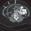
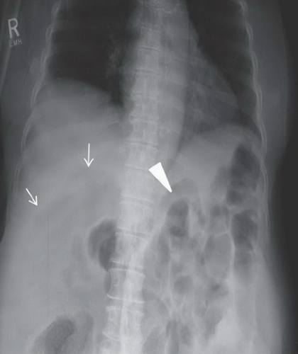 FIGURE 59A |
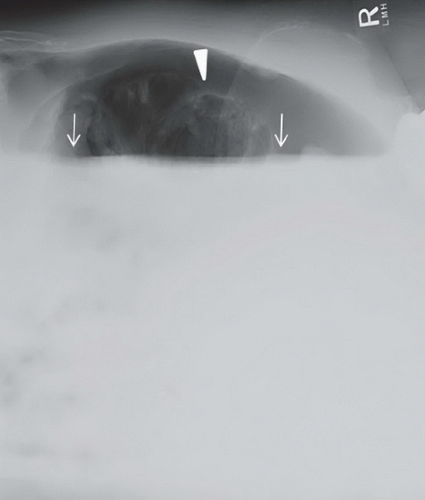 FIGURE 59B |
FINDINGS
Figure 59A: A supine abdominal radiograph demonstrates a large volume of free intraperitoneal air in the right hemiabdomen. The Rigler sign is identified with air present on both sides of the abdominal wall (arrowhead). The lucent liver sign is identified with increased lucency noted over the liver, which corresponds to free air anterior to the ventral surface of the liver (arrows). (Courtesy of Dr. Lauren Burke, Chapel Hill, NC, USA.) Figure 59B: A left lateral decubitus radiograph of the abdomen demonstrates a large volume of free intraperitoneal air in the nondependent abdomen between the abdominal wall and the liver, confirming pneumoperitoneum. The Rigler sign is again identified (arrowhead). There is an air-fluid level indicating fluid in the abdomen consistent with hydropneumoperitoneum (arrows). (Courtesy of Dr. Lauren Burke, Chapel Hill, NC, USA.)
DIFFERENTIAL DIAGNOSIS
Chilaiditi syndrome (colonic interposition between liver and hemidiaphragm), biliary or portal venous gas, fat within the subdiaphragmatic space or ligamentum teres, abscess, pneumatosis, gas within skin folds, properitoneal fat stripe, pneumoperitoneum.
DIAGNOSIS
Related posts:


Stay updated, free articles. Join our Telegram channel

Full access? Get Clinical Tree


