Acute Mesenteric Ischemia
Shaun R. Rybak
Ellie R. Lee
CLINICAL HISTORY
35-year-old female with acute onset abdominal pain. Initially, the abdominal pain significantly improved with pain medications and she was sent home. At home, the patient attempted to eat, but the abdominal pain returned.
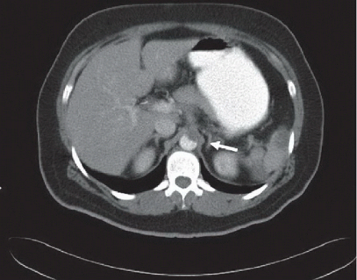 FIGURE 79A |
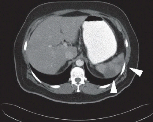 FIGURE 79B |
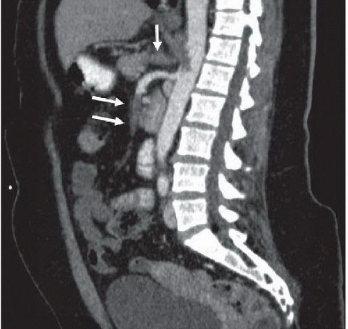 FIGURE 79C |
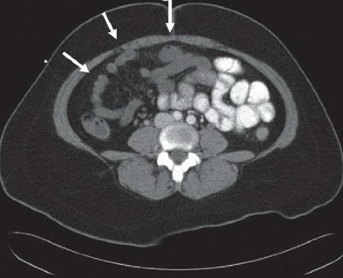 FIGURE 79D |
FINDINGS
Figure 79A: Axial contrast-enhanced CT image of the upper abdomen demonstrates thrombus within the anterior abdominal aorta (arrow). Figure 79B: Axial contrast-enhanced CT image of the upper abdomen demonstrates multiple peripheral wedge-shaped regions of hypodensity in the spleen (arrowheads), compatible with splenic infarcts. Figure 79C: Sagittal contrast-enhanced CT image of the abdominal aorta demonstrates filling defects in the enlarged proximal celiac artery and mid and distal superior mesenteric artery, consistent with acute thromboemboli (arrows). Figure 79D: Axial contrast-enhanced CT image of the abdomen demonstrates absent mural enhancement of the small bowel (arrows). The bowel was nonviable during surgery.
Related posts:


Stay updated, free articles. Join our Telegram channel

Full access? Get Clinical Tree


