Fig. 12.1
Risk of coronary events in relation to coronary artery calcium in the MESA study. The increase in coronary calcium score more than 100 was associated with more than sixfold increase in coronary events [39]
A meta-analysis by Pletcher et al. demonstrated that in comparison to CAC score of zero Agatston unit (AU), CAC score of (1–100 AU) has 2.1 relative risk for cardiac event including cardiac death, nonfatal myocardial infarction (MI), or stroke, while CAC score > 400AU has a much higher risk of cardiac event (relative risk 4.3) [40]. In another analysis, a CAC score of zero was associated with a very low risk of cardiac death or MI (annual event rate less than 0.4 %) [41]. In addition, many studies have shown that CAC provides incremental prognostic value over traditional risk factors [42] or biomarkers such as high sensitivity C-reactive protein [43]. However, the most important role of CAC remains its ability to reclassify patients within different risk categories as determined by the Framingham Risk Score (FRS) or even the newly described American College of Cardiology/American Heart Association risk estimator equation. Greenland et al. have showed that in the MESA cohort, the addition of CAC score to a prediction model based on traditional risk factors significantly improved the classification of risk and placed more individuals in the most extreme risk categories [44]. A total of 23 % of those who experienced events were reclassified to high risk, and an additional 13 % without events were reclassified to low risk. In the Heinz Nixdorf Recall study, CAC scoring reclassified 21.7 % and 30.6 % of the intermediate-risk group into the low- and high-risk groups, respectively, using CAC <100 and CAC >400 as cutoff values [45]. Recently, data from the MESA showed that nearly all risk score estimators overestimate the risk of patients by 25–115 % compared to calcium score using cardiac events as the endpoint [46].
CAC scoring has also an important role in the evaluation of patients with diabetes, a condition that is highly prevalent among patients referred to the nuclear labs. Several studies demonstrated that diabetic patients have a higher prevalence of CAC and a higher score compared to nondiabetic [47–49]. In a review of 10,377 asymptomatic individuals referred for CAC evaluation with a mean follow-up of 5 years, Raggi et al. found that CAC predicted all-cause mortality in diabetics [50]. In this cohort, the overall mortality was proportionate to the degree of calcification and at any given CAC score, the mortality was higher in diabetic compared to nondiabetic at any CAC score. In the same context, the absence of CAC was associated with low mortality irrespective of the presence of DM (Fig. 12.2).
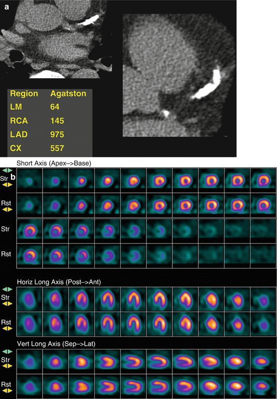

Fig. 12.2
(a) Coronary calcium score in a 71-year-old ex-smoker patient with chest pain and normal ECG. It shows severe coronary calcification in all three vessels with very high score of 1741 AU. (b) SPECT myocardial perfusion imaging: No significant perfusion defects are seen (other than diaphragmatic attenuation). The patient had severe three-vessel disease on coronary angiography and underwent coronary artery bypass surgery
However, the inherent difference between CAC and MPI should be kept in mind while utilizing both tests. While CAC is a measure of subclinical atherosclerotic burden, MPI evaluates the functional significance of coronary artery lesions. Looking at the same disease condition from different angles may provide more accurate diagnosis and assessment of the outcome. In the second part of this chapter, we will review the complimentary role of nuclear MPI and calcium score in patients referred to nuclear laboratories.
12.4 CAC Incremental Diagnostic Value
The addition of calcium score to nuclear MPI appears to add significant diagnostic value. CAC score could unmask significant CAD in patients with otherwise normal or low-risk MPI. Berman and colleagues evaluated 1195 subjects (51 % symptomatic) with both single-photon emission computed tomography (SPECT) and CAC scoring. Inducible ischemia was highly prevalent with high CAC and rarely seen when CAC <100 AU (<2 %) [51]. However, as shown in Fig. 12.3, many patients with normal SPECT MPI may have significant atherosclerosis which could be detected by CAC scoring [51].
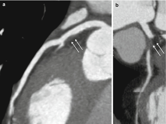

Fig. 12.3
Coronary CT angiography in a 70-year-old female with chest pain and normal ECG and cardiac markers. Coronary calcium score was zero. The patient has a non obstructive noncalcified plaque CAD in the LAD (a). The mid-RCA artery has a total occlusion with noncalcified plaque (b). Arrow indicates the location of the stenosis (a, b)
In a study of 77 intermediate-risk subjects referred for invasive angiography, CAC scoring and SPECT MPI were done [52]. A CAC score of 709 AU had high accuracy to detect significant CAD missed by SPECT MPI. This was also confirmed in another cohort of 864 asymptomatic patients [53] where a CAC score >1000 AU was able to detect significant coronary stenosis (≥50 %) on invasive angiography despite normal myocardial perfusion on SPECT [54]. This increased diagnostic accuracy is mostly related to the fact that a combined MPI-CAC study evaluates both anatomical and physiological aspects of the disease; thus allowing overcoming the limitations of physiological testing in patients where the anatomical burden of the disease is very significant.
12.5 Impact of Medical Therapy and Compliance
The identification of subclinical atherosclerosis by CAC can also help optimize the clinicians to optimize the medical therapy of patients with normal perfusion on MPI but higher atherosclerotic burden on CAC scoring. Nasir et al. found that only 59 % of subjects with CAC >400 AU and 65 % of subjects with CAC 100–399 AU did not qualify for pharmacotherapy by National Cholesterol Education Program (NCEP) Panel III guidelines leaving a significant number of patients with atherosclerosis without potentially lifesaving therapies [35]. Bybee et al. found that substantial number of patients with subclinical atherosclerosis and normal perfusion on PET were not receiving adequate therapy [55]. Only 41.5 % of patients with CAC 100–399 AU and 47.7 % of patients with CAC >400 AU were receiving statin with similar proportion of patients receiving aspirin. Increasing CAC score was associated with a higher chance of optimizing the medical therapy after PET result; the increasing CAC score was independently associated with initiation and optimization of medical therapy [55]. However, the prognostic value of this practice (adding intensive medical therapy to patients with normal perfusion and high CAC score) needs further evaluation and testing in a randomized controlled setting. Despite the proven utility of CAC in the diagnosis and prognosis of CAD, the exact score at which secondary preventive therapy should start is not well defined. The St. Francis Heart Study showed that asymptomatic patient with CAC >80th percentile did not benefit of daily atorvastatin therapy [56]. However, benefit with statin therapy was shown in a post hoc analysis of the same study in patients with CAC >80th percentiles and family history of CAD [57]. In an analysis of the MESA population who fulfilled the JUPITAR trial inclusion criteria, Blaha et al. reported that 5-year NNT was 24 and 19 for coronary heart disease and cardiovascular disease, respectively, when CAC >100 AU [58]. Therefore, although the exact CAC score to start statin therapy is not precisely defined, it is fair to say that statin therapy is reasonable to be started when CAC >100 AU [59]. Furthermore, it is important to note that the current guidelines recommend stress testing for asymptomatic patients with CAC >400 AU [60, 54].
12.6 Incremental Prognostic Value
The prognostic value of nuclear MPI has been extensively studied both with SPECT and PET imaging. While a normal exercise SPECT is associated with <1 % annual event rate of future cardiac event, an abnormal SPECT is associated with higher all-cause and cardiac mortality and cardiac events that are proportionate to the extent of abnormalities [61–63]. In addition, registry data suggested that nuclear MPI can be used to guide medical decisions. A reversible perfusion defect less than 10 % of the left ventricle would best be treated with medical therapy, while significant ischemia would best be treated with revascularization when feasible [64]. This approach is being tested in a large-scale National Institute of Health (NIH)-funded trial [65].
Several large-scale studies have examined the prognostic value of CAC over myocardial perfusion with SPECT. In 1126 patients (10 % diabetic) who underwent both CAC and SPECT MPI, Chang et al. found that all-cause mortality and nonfatal MI increased significantly with the severity of CAC [66]. CAC score >400 AU was associated with a 2.75-fold increase in all-cause mortality and nonfatal MI despite normal SPECT after a median follow-up duration of 7 years. Additionally, all-cause mortality and nonfatal MI exponentially increased with increasing severity of CAC in both normal and abnormal SPECT groups. Although the extent of ischemia on SPECT predicted hard cardiac event, adding CAC to the SPECT findings (normal/abnormal) further reclassified patients irrespective of the degree of ischemia on SPECT. Multiple smaller studies reported confirmed the above finding of the incremental prognostic value of CAC score over SPECT MPI [67, 68]. However, this added prognostic value may not be seen in short follow-up durations. A study of 1153 with a mean follow-up of 32 months showed that high CAC score failed to add incremental prognostic value in patients with normal perfusion on SPECT [69].
12.7 Addition of CAC to Positron Emission Tomography (PET)
The prognostic value of PET MPI has been demonstrated in several recent studies [70]. In a multicenter registry of 7061 patients referred for rubidium-82 stress PET, the extent and severity of perfusion abnormality were independent predictors of cardiac and all-cause mortalities [71]. Severely abnormal PET was associated with fivefold increase in cardiac mortality, and perfusion PET was able to reclassify nearly 12 % of patients. In addition, noninvasive coronary flow reserve adds incremental prognostic value to perfusion abnormalities on PET [72, 73].
In concordance with SPECT literature, a study of 695 intermediate-risk patients who underwent rubidium-82 PET imaging showed that an abnormal PET scan was seen more often with increasing CAC score [74]. The frequency of abnormal scans among patients with a CAC score > or = 400 was higher than that in patients with a CAC score of 1–399 (48.5 % versus 21.7 %, P <0.001). However, the frequency of ischemia among patients with no CAC was 16.0 %, and its absence only afforded a negative predictive value of 84.0 %. Among patients with normal PET myocardial perfusion imaging, the annualized event rate in patients with no CAC was lower than in those with a CAC score > or = 1000 (2.6 % versus 12.3 %, respectively). Likewise, in patients with ischemia on PET myocardial perfusion imaging, the annualized event rate in those with no CAC was lower than among patients with a CAC score > or = 1000 (8.2 % versus 22.1 %) (Fig. 12.4).
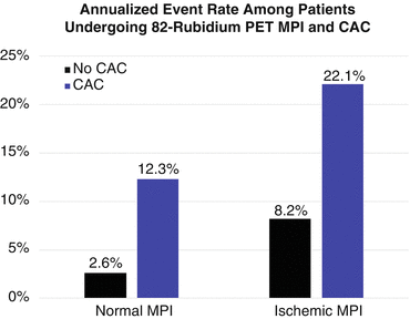

Fig. 12.4
Event rate among patients undergoing both 82-rubidium PET MPI and CAC. Among patients with normal PET myocardial perfusion imaging, the annualized event rate in patients with no CAC was lower than in those with a CAC score ≥ 1,000 (2.6 % versus 12.3 %, respectively). Likewise, in patients with ischemia on PET myocardial perfusion imaging, the annualized event rate in those with no CAC was lower than among patients with a CAC score ≥ 1,000 (8.2 % versus 22.1 %)
This was also seen in patients referred to our laboratory. With very high prevalence of diabetes mellitus (55 %), hypercholesterolemia (90 %), and hypertension (87 %), the prevalence of perfusion defects increased with increasing CAC score. While nearly 54 % of patients with CAC >400 AU had an abnormal scan [75], the absence of CAC excluded ischemia in 206 intermediate-risk subjects with a negative predictive value (NPV) of 99 % (Fig. 12.5) [76]. These findings support the notion that CAC is predictive of abnormal myocardial perfusion on PET and thus may be used to improve the diagnostic accuracy of PET MPI.
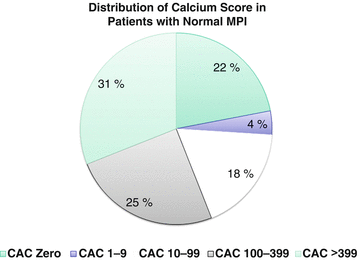

Fig. 12.5
Distribution of coronary calcium score in a patients with normal myocardial perfusion imaging. Patients with normal myocardial perfusion imaging have a wide range of coronary calcium. Nearly one third of the patients have high calcium score of more than 400 [51]
Since most PET systems are hybrid machines including both PET and CT used primarily for attenuation correction, CAC score can be easily performed in the same setting. The interplay between CAC and coronary flow reserve (CFR) measured on PET was investigated in 901 patients with a mean follow-up of 1.53 years. The annual major adverse cardiac event (MACE), including cardiac death, nonfatal MI, revascularization, and admission for heart failure, increased with CFR <2 [77]. Although there was no direct association between CAC and short-term MACE in this study, CFR <2 occurred at higher frequency with increasing CAC score. Moreover, there was a significant association between low CFR (<2) and annual MACE across all levels of CAC [77]. The findings of this study may suggest that CAC although reflective of overall atherosclerotic burden is likely to represent a late manifestation of atherosclerosis than reflecting the activity of the disease. CAC may be predicative of late events, while CFR and myocardial ischemia could be predictive of short-term events.
Finally, evaluating CAC allows for measurement of other potential high-risk markers. There is some data to suggest that epicardial fat is associated with presence and outcome of CAD [78, 79]. It can be easily measured on the gated non-contrast CT used for CAC scoring. It has been associated with ischemia on SPECT MPI with odd ratio of 2.68 in patients without known CAD [80]. Measurement of pericardial fat may help in the decision making of obscure cases of SPECT and may help further in stratification of the patients in general. In addition, other vascular and cardiac nonvascular calcifications can be measured, many of which add independent prognostic value. However, further data is needed to confirm the added role of epicardial fat and noncoronary cardiac calcifications.
12.8 Practical Considerations
Despite the clearly demonstrated incremental diagnostic and prognostic value of coronary calcium score in different patient populations, some practical considerations need to be taken into account. Given the fact that CAC and perfusion imaging study are two different distinct processes, it would be expected to have clinical scenarios where the two tests yield discrepant results. The impact of these discrepant results on the patient acute management is not well studied. Management decision should take into account the patients’ baseline risk and symptomatic status as well as baseline coronary disease status. Since coronary calcium score has limited role in patients with known coronary disease (prior angioplasty or bypass surgery), these patients need to be primarily studied by perfusion imaging. On the other hand, one would expect a very low prevalence of perfusion defects in patients with zero calcium score (<5 %). Artifacts have to be thought of in this patient population of zero calcium score including misregistration artifacts. Untimely revascularization decisions should be made based on the presence of perfusion defects and the extent of myocardial ischemia. Thus, patients with normal perfusion and patients with high calcium score should not be subjected to coronary angiography unless balanced ischemia is suspected. On the other hand, patients with a true perfusion defect should not be deprived from revascularization just because their calcium score is low.
12.9 Conclusion
In conclusion, CAC scoring is a robust and effective tool for the detection of anatomical atherosclerosis, while myocardial perfusion imaging is a superb and validated technique to detect myocardial ischemia and prognosticate patients. While CAC and MPI evaluate two deferent aspects of CAD, anatomy versus physiology, it is likely that in a moderate- or high-risk population, adding CAC measurement to myocardial perfusion imaging with PET and SPECT imaging would give an incremental diagnostic and prognostic value. It will also aid in optimizing the medical therapy and risk stratification of subjects. However, revascularization decisions should be solely made based on the ischemic evaluation rather than the anatomic manifestation of atherosclerosis.
References
1.
Go AS, Mozaffarian D, Roger VL, et al. American Heart Association Statistics C, Stroke Statistics S. Heart disease and stroke statistics–2014 update: a report from the american heart association. Circulation. 2014;129:e28–e292.
2.
Holly TA, Abbott BG, Al-Mallah M, et al. American Society of Nuclear C. Single photon-emission computed tomography. J Nucl Cardiol. 2010;17:941–73.
3.
4.
5.
De Lorenzo A, Lima RS, Siqueira-Filho AG, Pantoja MR. Prevalence and prognostic value of perfusion defects detected by stress technetium-99m sestamibi myocardial perfusion single-photon emission computed tomography in asymptomatic patients with diabetes mellitus and no known coronary artery disease. Am J Cardiol. 2002;90:827–32.PubMedCrossRef
6.
7.
Iskandrian AS, Chae SC, Heo J, Stanberry CD, Wasserleben V, Cave V. Independent and incremental prognostic value of exercise single-photon emission computed tomographic (SPECT) thallium imaging in coronary artery disease. J Am Coll Cardiol. 1993;22:665–70.PubMedCrossRef
Stay updated, free articles. Join our Telegram channel

Full access? Get Clinical Tree


