Adrenal Hemorrhage
Kavya E. Reddy
Ellie R. Lee
CLINICAL HISTORY
18-year-old female in a trauma—motor vehicle versus pedestrian.
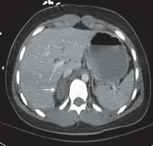 FIGURE 7A |
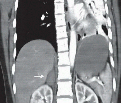 FIGURE 7B |
FINDINGS
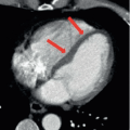
Figures 7A and 7B: Axial and coronal contrast-enhanced CT images of the abdomen demonstrate an enlarged, dense hematoma in the right adrenal gland, measuring 56 HU, with mild adjacent stranding (arrow). Normal left adrenal gland. Left pneumothorax and collapse of the left lung identified on the coronal image.
DIFFERENTIAL DIAGNOSES
Adrenal hemorrhage, adrenal adenoma, adrenal carcinoma, adrenal metastasis, adrenal lymphoma, infection of the adrenal gland (TB or fungal).
DIAGNOSIS
Related posts:

Stay updated, free articles. Join our Telegram channel

Full access? Get Clinical Tree


