Fig. 17.1
Illustration of electron-tracking based Compton imaging. Without the knowledge of the electron scatter direction, the direction of the incident gamma can only be restricted to a cone surface. By measuring the initial electron trajectory the cone can be reduced to an arc reducing the background in the reconstructed gamma-ray image. The resolution, i.e. the extension of the arc depends on the resolution of the electron track reconstruction. The intrinsic width of the cone and arc depends on the energy and position resolution for individual gamma-ray interaction of the instrument and ultimately, the uncertainty due to the intrinsic momentum of the Compton-scattered electron
Fig. 17.2
Illustration of potential gain by employing electron-tracking based Compton imaging in comparison with conventional Compton imaging. Simple cone backprojection is used assuming an angular electron track reconstruction resolution of 30
We were able, for the first time, to demonstrate electron-tracking based Compton-imaging in a solid-state detector [5, 6]. While this concept has been shown before in gas-based instruments, these detectors are characterized by very low interaction efficiency and low energy resolution, significantly limiting the achievable gamma-ray imaging sensitivity [7]. The scientific CCDs we are using are fully depleted Si devices with a thickness of 650 um and a pixel pitch of 10.5 μm [8]. These performance characteristics are well suited for tracking electrons above about 150 keV. We have demonstrated the ability to reconstruct gamma rays with a arc opening angle of about 30–60°, depending on incident angle and deposited energy [9].
While the accurate and efficient measurement of the initial direction of the scattered electron represents a significant challenge for the detection instrument, the conversion of the measured energy and position signals into an energy and direction of the incident gamma rays requires sophisticated and newly developed algorithms. For example, we have developed algorithms to extract the initial three-dimensional scatter direction of the electron by only observing the two-dimensional energy loss distributions in the used CCD [10]. Furthermore, we have developed a new algorithm that allows us to analytically reconstruct the incident gamma-ray energy and angle by just using the initial electron scatter direction and the energy deposited by the Compton scattered electron, i.e. by only measuring the partial energy of the incident gamma ray [11]. This realization potentially allows us to reconsider inherently inefficient gas detectors, as only one interaction is required. Figure 17.3 illustrates the results of the reconstruction of a Cs-137 source characterized by a gamma-ray energy of 662 keV.
Fig. 17.3
Reconstruction of gamma-ray momentum, i.e. energy and incident direction by using the measured initial electron scatter direction and electron energy in a CCD system. Top left is the measured electron energy in the CCD reflecting the energy deposited by the Compton scattered electron. Note that the full energy of the gamma ray is not visible as only the energy of the Compton-scattered electron is deposited. Top right: Reconstructed gamma-ray momentum. Bottom left: Projection of momentum on energy axis illustrating the reconstruction of the incident, full gamma-ray energy. Bottom right: Reconstructed incident direction of gamma rays
17.2.2 3D Volumetric Imaging
Complementary to μm-resolution and small-scale instruments used to develop a new generation of gamma-ray imaging instruments as discussed above, we are pursuing the development of medium-sized and hand-portable gamma-ray detection and imaging systems in combination with contextual sensors. These combined 3D volumetric imaging systems enable the simultaneous reconstruction of the 3D environment or scene and the fusion of gamma-ray images with this 3D world. This 3D fusion goes beyond the already developed overlay of visual images and gamma-ray images in two-dimension by fully integrating multi-sensor information in three dimensions. The advantages are the significantly increased efficiency, accuracy, and contrast in the measurement of the gamma-ray information, the possibility for quantification of gamma-ray sources, and the increased speed in the gamma-ray reconstruction. As development platform, we are using the second generation Compact Compton Imager (CCI-2) and the High-Efficiency Multimode Imager (HEMI) [12–14]. Both systems are shown in Fig. 17.4.
Fig. 17.4
Left: Second generation Compact Compton Imager consisting of 2 DSSD HPGe detectors mounted in a cryostat with contextual sensors, two panoramic video cameras and a Lidar scanner. Right: The hand-portable High-Efficiency Multimode Imager consisting of arrays of CPG CZT detectors to enable coded aperture and Compton imaging, simultaneously
CCI-2 consists of two high-purity Ge (HPGe) detectors implemented in a double-sided strip detector (DSSD) configuration. Each detector has a thickness of 15 mm and 38 orthogonal strips with a strip pitch of 2 mm. Digital signal processing is used to obtain the position of individual gamma-ray interactions to about 1 mm in all three dimensions. This position resolution combined with the energy resolution of about 2 keV for each strip provides an angular resolution of about 4° when operated in Compton imaging mode. The angular resolution depends on the energy and data cuts implemented reflecting the trade-off between efficiency and angular resolution. The system is mounted on a cart providing support for the fully digital data acquisition system to read out the 152 strips, the power supplies, and the liquid nitrogen dewar to provide the necessary cooling for the HPGe detectors. While this system is too heavy for easy deployment and represents an efficient platform for the development of detection and data fusion concepts. A more compact and hand-portable, DSSD HPGe based system is commercially available, but with reduced position resolution [15]. HEMI consists of 96 CdZnTe (CZT) detector implemented in a coplanar grid (CPG) configuration [16]. The individual, 1 cm3 big CZT detectors are arranged in two parallel planes with 32 detectors in the front plane and a fully populated plane of 64 detectors arrange in a 8 × 8 array. The front plane of active detectors serves as coded aperture for low gamma-ray energies and as scatter plane for Compton imaging for higher gamma-ray energies. The total active detector mass is 1.2 lbs with the whole instrument weighing about 8 lbs and is therefore easily portable. In comparison to the DSSD HPGe the energy resolution is degraded and the position resolution is limited to 1 cm3 resulting in an angular resolution of about 10°at 662 keV. Both systems have been combined with contextual sensors such as high-resolution Ladybug video cameras and 32 beam rotating Lidar systems or Microsoft Kinect© sensors.
Figure 17.5 illustrates the reconstruction and fusion of objects and images in 3D. By moving the cart or walking with HEMI through the scene, it is possible to reconstruct a point cloud representing surfaces of objects in 3D and to use these point clouds to increment the gamma-ray image.
Fig. 17.5
Illustration of 3D volumetric imaging. 3D point clouds are registered using a Microsoft Kinect sensor mounted to the CCI2 gamma-ray imaging systems. By moving the cart through the cluttered scene, the 3D point cloud is obtained and subsequently used to reconstruct the gamma-ray image in 3D. The red line indicates the trajectory of the instrument, the blue arrows represent the gamma-ray scatter directions. These events have been used to reconstruct the location of the Cs-137 source. The total acquisition time was less than 90 sec. while the cart was moved over a distance of about 12 ft
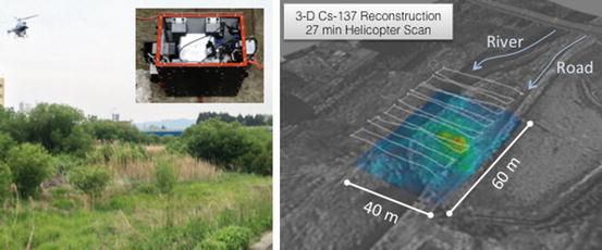
In collaboration with JAEA, HEMI has also been deployed to Fukushima to evaluate contamination mapping capabilities when mounted on a RMAX unmanned and remotely controlled helicopter. The dimensions and the weight of HEMI are well suited for these demonstrations. The HEMI instrument itself was packaged in an environmental box that allowed an operation completely independently from the helicopter operation. The whole package included a video camera, a GPS/IMU system, thermal stabilization, data storage, batteries, a control unit, and HEMI. The video information is used to reconstruct three-dimensional surfaces on the ground stereoscopically. These surfaces are then used to project the gamma-ray images onto as shown in Fig. 17.6. Although these 3D surfaces improve the reconstruction, it still provides limited reconstruction capabilities. This is due to the fact that the surfaces are typically the surfaces of vegetation and not the source of the radiation which is located beneath the top surfaces, e.g. in the soil or the whole plant. More work remains to be done to increase the quantitative reconstruction capabilities in the complex environments of Fukushima.
 To Pursue an Independent Nuclear Deterrent or Not? Japan’s and South Korea’s Nuclear Decision Making Models
Neutralizing Radicalized Threat Networks, Disrupting WMD Illicit Traffickers and Targeting Corrupt Facilitators
A Holistic Approach to Radiological Terrorism
To Pursue an Independent Nuclear Deterrent or Not? Japan’s and South Korea’s Nuclear Decision Making Models
Neutralizing Radicalized Threat Networks, Disrupting WMD Illicit Traffickers and Targeting Corrupt Facilitators
A Holistic Approach to Radiological Terrorism
 Dynamics of International Nuclear Safety: Post-Fukushima Developments in Regulatory Oversight and Filtered Vent Technology in Six Nuclear Countries
 Current Activities of the European Union in Fighting CBRN Terrorism Worldwide
 Will the Implementation of Small Modular Reactors Affect Our Response Protocols to Nuclear and Radiological Threats?


Related posts:
To Pursue an Independent Nuclear Deterrent or Not? Japan’s and South Korea’s Nuclear Decision Making Models
Neutralizing Radicalized Threat Networks, Disrupting WMD Illicit Traffickers and Targeting Corrupt Facilitators
A Holistic Approach to Radiological Terrorism
Dynamics of International Nuclear Safety: Post-Fukushima Developments in Regulatory Oversight and Filtered Vent Technology in Six Nuclear Countries
Current Activities of the European Union in Fighting CBRN Terrorism Worldwide
Will the Implementation of Small Modular Reactors Affect Our Response Protocols to Nuclear and Radiological Threats?
Stay updated, free articles. Join our Telegram channel
Full access? Get Clinical Tree
