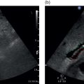
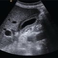
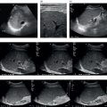
Adrian K.P. Lim1,2, James P.F. Burn1, and Caroline Ewertsen3 1 Department of Imaging, Imperial College Healthcare NHS Trust, London, UK 2 Department of Metabolism, Digestion and Reproduction, Imperial College London, UK 3 Department of Radiology, Rigshospitalet, Copenhagen, Denmark Ultrasound technology continually improves in leaps and bounds and when first introduced, it was primarily to assess anatomical structures and detect focal lesions. The development of Doppler and subsequently contrast ultrasound added yet another dimension to ultrasonic assessment of the liver, and the current state‐of‐the‐art technologies also employ texture analysis and elastography, with a multiparametric approach to non‐invasive liver assessment in chronic hepatic disease. This chapter is a summary of current available technologies (accurate at time of publication), some of which are still undergoing trials to evaluate their efficacy. Optimum image resolution has been the paramount goal of ultrasound manufacturers, where the detection of focal liver lesions and also texture appreciation rival those of other imaging modalities, namely computed tomography (CT) and magnetic resonance imaging (MRI), where sub‐centimetre lesions can be better characterised with ultrasound. The spatial and temporal resolution is improved with better probe technologies, where the penumbra of the frequency range of the probe is increased, covering ranges between 2 and 9 mHz rather than 1–5 mHz previously. The ability to generate thinner ultrasound beams also provides improved image homogeneity and image processing algorithms remove noise and artefact, giving a ‘cleaner’ image. One constant limiting factor has also been body habitus, where an increase in body mass index (BMI) significantly reduces ultrasound penetration and resolution. There are now lower‐frequency probes allowing visualisation up to a 30 cm depth, but of course with a trade‐off of poorer image quality than with more conventional probes. However, some imaging is better than none, particularly in populations where obesity is becoming an ever‐increasing problem. Microvascular flow imaging (MFI) is the general terminology to encompass the more sensitive Doppler offerings, where clever algorithms allow the depiction of slow‐flowing vessels separate from noise. The common acronyms include superb microvascular imaging (SMI, Canon Medical Systems, Tochigi, Japan), micro‐flow imaging (MFI, Philips Medical Systems, Eindhoven, Netherlands), or just microvascular imaging (MVI, Samsung Medical Systems, Kyonggi, South Korea). GE Healthcare (Chicago, IL, USA) has in addition to microvascular flow Doppler technology, a version that utilises B‐mode rather than Doppler technology and is termed B‐flow. MFI applies an advanced Doppler algorithm and unique wall filters to identify, isolate, and eliminate clutter. The result is preservation of low‐velocity flow signals, allowing representation of microvessel blood flow to be depicted. This is achieved at low mechanical index, high frame rates (enabled with plane wave imaging), and without the need for contrast agents. The end result is a Doppler technique that has significantly improved sensitivity and spatial resolution, in comparison to standard colour Doppler/power Doppler ultrasound [1–4]. The visualisation is much refined (Figure 15.1), but is technically not seen down to micrometres and the vessels depicted are in the range of 1 mm or so. Nonetheless, this adds to the diagnostic capability and can sometimes obviate the need for contrast enhancement, particularly in the correct clinical setting (Figures 15.2 and 15.3). Figure 15.1 (Video 15.1) This shows a thrombosed portal vein with numerous collaterals. The right‐hand image is the monochrome version of superb microvascular imaging (SMI), Canon Medical Systems’ version of microvascular flow imaging. Note the superb resolution of the small vessels and collaterals, which are typically not shown with colour Doppler. Credit: A. Wilson, A.K.P. Lim., (2022), ELSEVIER, Licenced under CC BY 4.0. Figure 15.2 (Video 15.2) There is an exophytic focal lesion on a background of liver cirrhosis. Note the nodular edge and ascites within segment VI of this liver. The right‐hand image is with superb microvascular imaging (SMI) turned on. Note how clearly the abnormal haphazard neoangiogenic vessels are displayed without the use of contrast. This microvascular pattern clearly denotes a hepatocellular carcinoma. Credit: A. Wilson, A.K.P. Lim., (2022), ELSEVIER, Licenced under CC BY 4.0. Figure 15.3 (Video 15.3) This was an incidental finding in a young female patient with no medical history of note. There was suggestion of a subtle hypoechoic lesion in the liver and on turning on monochrome superb microvascular imaging (SMI; right‐hand image), the stellate, spoke–wheel pattern of this vascular lesion is clearly depicted. There has been no injection of contrast and, given the lack of a history of malignancy or chronic liver disease, it can safely be diagnosed as focal nodular hyperplasia (FNH
15
Advancing Ultrasound Technologies
Ultrasound B‐Mode/Greyscale Imaging
Doppler Technologies


Related posts:
![]()
Stay updated, free articles. Join our Telegram channel


Full access? Get Clinical Tree


