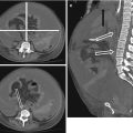
Fig. 17.1
Space-occupying MCA-infarction before (a) and after osteoclastic trepanation (b); space-occupying PICA-infarction at onset (c) and after suboccipital craniectomy (d)
Spontaneous intra-cerebral hemorrhage is the cause of 10–15 % of strokes. Compared to ischemic strokes it carries a poorer prognosis, and the 3-month mortality is reported to be up to 40 %. Neurological symptoms depend on the site and size of the bleeding. Intracranial hypertension occurs when the hemorrhage exceeds a critical volume. The clinical signs are non-specific and urgent imaging with CT or MRI is required when the index of suspicion is high (Fig. 17.2a–c).
In case of subarachnoid hemorrhage cerebral aneurysms or vascular malformations must be excluded (Fig. 17.2d–f). CT angiography (CTA) or MR angiography (MRA) should be considered as first diagnostic step. However, invasive angiography is still the gold standard in cases of subarachnoid hemorrhage, as CTA or MRA may miss small aneurysms of the basal cerebral arteries.
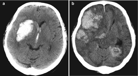
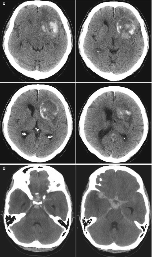
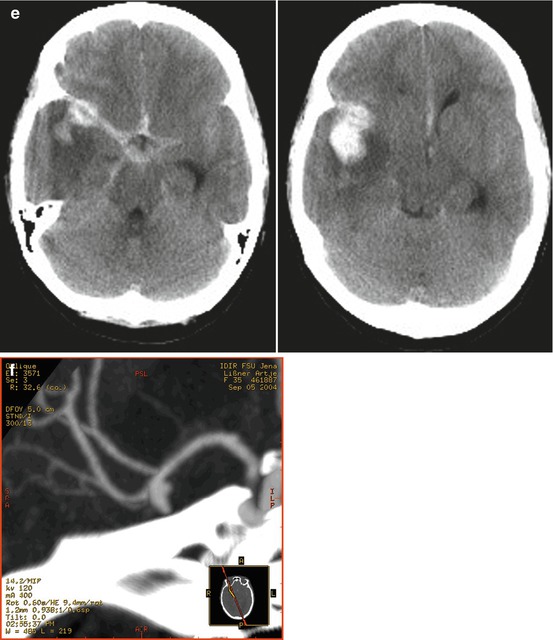


Fig. 17.2
Unenhanced CT images of spontaneous intracerebral haemorrhage. (a) Due to hypertension; (b) due to a coagulation disorder; (c) tumor hemorrhage (nativ and with contrast media); (d, e) subarachnoid hemorrhage due to an aneurysm of the right middle cerebral artery; (f) CT Angiogram showing the aneurysm
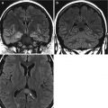
Atypical spontaneous intracerebral hemorrhage can be caused by cerebral sinus thrombosis. The bleeding is localized near the occluded vein. Venous angiography with contrast CT or MR-angiography is the imaging modality of choice to detect sinus thrombosis. Catheter angiography is only required in indeterminate cases.
Meningitis, Encephalitis and Sequelae from Infective Endocarditis
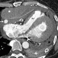
Infections of the meninges or the CNS due to bacterial, viral, fungal or parasitic microorganisms may present with broad array of unspecific symptoms including fever, altered level of consciousness and focal neurological deficits. These conditions are potentially life-threatening, but if the diagnosis is made early successful antimicrobial treatment is often possible. Infections of the central nervous system are described in more detail in Chap. 15.
Stay updated, free articles. Join our Telegram channel

Full access? Get Clinical Tree


