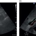
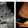
Adrian K.P. Lim1,2, Chris J. Harvey1, and Matteo Rosselli3,4 1 Department of Imaging, Imperial College Healthcare NHS Trust, London, UK 2 Department of Metabolism, Digestion and Reproduction, Imperial College London, UK 3 Department of Internal Medicine, San Giuseppe Hospital, USL Toscana Centro, Empoli, Italy 4 Division of Medicine, Institute for Liver and Digestive Health, University College London, Royal Free Hospital, London, UK Microbubbles form the basis of ultrasound contrast agents (UCAs), where one of the first agents used was agitated saline. Following on from this was an agent that consisted of tiny microbubbles of air stabilised with a surfactant layer, which had the trade name Levovist (Schering, Berlin, Germany). It had a stability of only 30 minutes once constituted. However, it has now been superseded by more stable agents. These typically contain a perfluorocarbon gas and a phospholipid outer shell for improved stability, and can be used for diagnostic purposes for up to six hours once they have been mixed. Many of the agents are constituted by mixing a prepared syringe of a solvent into a vacuum‐sealed vial containing a powder and the perfluorocarbon gas. The mixing and shaking of the two produces microbubbles that range in size, typically between 1 and 10 μm. The most commonly used agent in Europe is SonoVue, but there are several others licensed for clinical use. Currently there are four agents that are available internationally: The approval of these agents varies throughout the world along with the approved indications, and is dependent on the regulatory agency of the country of use. UCAs remain in the intravascular space and do not diffuse out into the extracellular fluid. The advantage of their being confined to the vascular compartment is that this provides improved delineation of the microvasculature, better than the iodinated or gadolinium‐based agents used in computed tomography (CT) and magnetic resonance (MR) imaging, respectively [1]. Interestingly, although there is no renal excretion unlike CT/MR agents, a couple of these agents where either phagocytosis by Kuppfer cells has been reported or they remain stationary for a period of time within the sinusoidal spaces of the liver [2]. These days there are target‐specific microbubbles such as those conjugated with an anti‐vascular endothelial growth factor receptor‐2 monoclonal antibody. These could potentially help image the vascularisation of a tumour. Much of the work has been on animal models, but some are currently being trialled for prostate tumours [3, 4]. When UCAs are insonated they contract, expand, and contract, producing oscillations that are asymmetrical in diameter. These generate harmonic signals and it is fortuitous that microbubbles resonate at the lower frequency range used in diagnostic imaging. The propagation of the ultrasound wave also demonstrates similar non‐linear behaviour in biological tissue, but to a lesser extent than with UCAs. It is this property that ultrasound manufacturers have utilised to tease out the signal difference between tissue and microbubbles. Many scanners have a contrast mode, which utilises a pulse inversion or pulse modulation technique or sometimes a combination, to amplify the signals by UCAs while cancelling those from tissue. These are low power modes, typically one‐tenth of that used for the greyscale image. At higher powers, the microbubbles will burst and could lead to a loss of signal, with apparent non‐enhancement or washout. Each manufacturer has its own acronym for contrast mode, usually with ‘harmonic imaging’ in the name. Some have simplified this to just ‘contrast’ and many have also ensured that all the buttons necessary to perform the contrast study are ergonomically positioned close together. They also ensure that adequate cineloops of at least 60 seconds or more are enabled for storage. The preferred on‐screen display is a split‐screen set‐up, where one half depicts the microbubble and the other side is the greyscale localiser (Figure 4.1). The option of having just the microbubble signature display is also possible, as is an overlay of this mode on the greyscale image. The microbubble display is usually a bright, distinct colour to enable easy visualisation, typically with a gold/chrome hue. On some scanners a dual cursor also allows the more accurate localisation of a small lesion, ensuring that the enhancement pattern can be accurately assessed (Figure 4.2).
4
An Introduction to Contrast‐Enhanced Ultrasound
Ultrasound Contrast Agents
Pharmacokinetics
Interaction with Ultrasound Waves
Scanning Modes
Stay updated, free articles. Join our Telegram channel

Full access? Get Clinical Tree


