and Lori Ann Lewis1
(1)
Clinical Research Center of North Texas, Denton, TX, USA
Abstract
Osteoporosis is not the only disease in which bone densitometry is used in diagnosis and management, but it is the most important, from the standpoint of the prevalence of the disease itself and the number of individuals referred for testing. It is not the responsibility of the technologist to discuss disease processes with patients referred for testing. In fact, some physicians would consider this intrusive and inappropriate. Nevertheless, the setting in which densitometry is usually performed and the interaction between the technologist and patient is conducive to patients asking questions of the technologist about osteoporosis. In these circumstances, it would be inappropriate for a technologist to fail to respond within reason or appear to be uninformed. Knowledge of the disease process and the approved therapies for osteoporosis should be part of the densitometry technologist’s education. In any discussion with patients, however, it should also be emphasized that the patient’s physician is the final authority on the interpretation of bone density results and the need for prescription or nonprescription interventions to prevent or treat osteoporosis.
Osteoporosis is not the only disease in which bone densitometry is used in diagnosis and management, but it is the most important, from the standpoint of the prevalence of the disease itself and the number of individuals referred for testing. It is not the responsibility of the technologist to discuss disease processes with patients referred for testing. In fact, some physicians would consider this intrusive and inappropriate. Nevertheless, the setting in which densitometry is usually performed and the interaction between the technologist and patient is conducive to patients asking questions of the technologist about osteoporosis. In these circumstances, it would be inappropriate for a technologist to fail to respond within reason or appear to be uninformed. Knowledge of the disease process and the approved therapies for osteoporosis should be part of the densitometry technologist’s education. In any discussion with patients, however, it should also be emphasized that the patient’s physician is the final authority on the interpretation of bone density results and the need for prescription or nonprescription interventions to prevent or treat osteoporosis.
The Definition of Osteoporosis
The 1991 and 1993 Consensus Development Conferences
The most widely accepted formal definition of osteoporosis was originally proposed in 1991 and reaffirmed in 1993 at consensus development conferences sponsored by the National Osteoporosis Foundation, the European Foundation for Osteoporosis and Bone Disease, and the National Institute of Arthritis and Musculoskeletal and Skin Diseases. At those conferences, osteoporosis was defined as “a systemic skeletal disease, characterized by low bone mass and microarchitectural deterioration of bone tissue with a consequent increase in bone fragility and susceptibility to fracture” [1, 2]. This definition of osteoporosis was a departure in many respects from previous definitions of the disease. Prior to 1991, osteoporosis was often described as an “age-related” disorder, which implied that the inevitability of advancing age alone was reason to develop the disease. This also implied an inability to prevent or even successfully treat osteoporosis. In the 1991 and 1993 consensus conference definitions, there is no longer any mention of aging as a causative factor.
Some definitions of osteoporosis also required that a fracture be present before the disease could be said to exist. The 1991 and 1993 consensus conference definition does not require the presence of a fracture. The definition requires only that the skeleton be sufficiently fragile that an individual is at increased risk for fracture. This approach separates the undesirable outcome of a fragile skeleton, fracture, from the disease process itself. This is similar to the approaches taken with hypertension and hypercholesterolemia. For example, the disease hypertension is based on the finding of an increased blood pressure, a quantity that is measured clinically. Once the blood pressure exceeds a certain limit, hypertension is said to exist. Having hypertension places the individual at increased risk for a stroke, although hypertension is not the only cause of stoke. The undesirable outcome of hypertension, then, is a cerebrovascular accident or stoke. The presence of a stroke, however, is not required before it can be said that the disease hypertension exists. The same is true with hypercholesterolemia. This diagnosis is based on the finding of increased levels of cholesterol in the blood, a quantity that is measured clinically. The undesirable outcome of hypercholesterolemia is myocardial infarction or heart attack. It is not necessary for a heart attack to have occurred, however, before the diagnosis of hypercholesterolemia is made.
Hypertension and hypercholesterolemia are both diseases that are based on finding abnormal values of quantities that can be measured clinically, blood pressure and cholesterol. Like these diseases, the 1991 and 1993 consensus conference definitions of osteoporosis suggested that osteoporosis could, at least in part, be defined on the basis of a quantity that could be measured such as the bone mass or density. The clinical measurement of microarchitectural deterioration of bone tissue in vivo remains difficult even today. But the bone mass or density can be readily measured by any one of several different techniques. It only remained to define the level of bone density that resulted in an increased risk for fracture to complete a clinically useful definition of osteoporosis.
The 1994 World Health Organization Criteria for Diagnosis of Osteoporosis
The World Health Organization (WHO) criteria for the diagnosis of osteoporosis based on the measurement of bone density were published in 1994 [3]. At the time the criteria were developed, the WHO was attempting to devise criteria that would allow them to estimate the prevalence or percentage of individuals in different countries who might have osteoporosis. In order to do this, some common objective definition of osteoporosis was required. The WHO was actually not attempting to specify a level of bone density that would be used clinically in individuals to diagnose osteoporosis.
The levels of bone density that were ultimately chosen by the WHO were based on reviewing the medical literature that was available at the time. After considering several different approaches to establishing the level of bone density that would be called osteoporosis, the WHO stated that a bone mass or bone density that was 2.5 standard deviations (SD)1 or more below the average peak bone mass or density of the young adult was sufficiently low to be called osteoporosis [3]. This was based on the finding that the percentage of women in the United States and Great Britain who were thought to have a bone density this low at the hip or at the hip, spine, and forearm combined was very similar to the lifetime risk of hip fracture and the lifetime global fracture risk.2 The bone mass or bone density was considered normal if it was not more than 1 SD below the average peak bone density of the young adult. Bone mineral densities that were more than 1 but less than 2.5 SD below the average young adult value were called osteopenic or simply low. A fourth category, called severe or established osteoporosis, referred to individuals who had bone densities that were 2.5 SD or more below the average for a young adult and who also had a fracture. These criteria are summarized in Table 9-1 and again in Appendix B for easy reference. The WHO did not restrict the application of these criteria to measurements at any particular skeletal site while noting that measurements at different sites could result in different diagnoses. The original WHO criteria were given as the number of SDs below the average peak bone mass or density for each diagnostic category. These criteria can readily be converted to T-scores as shown in Table 9-1 since the definition of the T-score in bone densitometry indicates the number of SDs above or below the average peak bone mass or density.3
Table 9-1
The World Health Organization Criteria for the Diagnosis of Osteoporosis Based on the Measurement of Bone Density
Diagnosis | Bone density criteria | T-score criteria |
|---|---|---|
Normal | Not more than 1 SD below the average peak young adult value | Better than or equal to −1 |
Osteopenia (low bone mass) | More than 1 but not yet 2.5 SD below the average peak young adult value | Poorer than −1 but better than −2.5 |
Osteoporosis | 2.5 SD or more below the average peak young adult value | −2.5 or poorer |
Severe (established) osteoporosis | 2.5 SD or more below the average peak young adult value + a fracture | −2.5 or poorer + a fracture |
It should be noted again that the WHO was not attempting to establish diagnostic criteria for the clinician to use in diagnosing individual patients. With the increasing usage of bone densitometry, however, and the consensus conference definitions of osteoporosis that included a finding of low bone mass without an objective level being specified, clinicians understandably began to apply the WHO criteria to individual patients. The WHO criteria were based on information that was relevant only to Caucasian women. Strictly speaking, then, the WHO criteria should be applied only to Caucasian women. In addition, they were most relevant to postmenopausal Caucasian women. The WHO did note in 1994 that in the absence of other criteria, it might not be inappropriate to apply the WHO criteria to mature Caucasian men. There is increasing consensus that the WHO criteria are appropriate for mature men and postmenopausal women of other races. There is little disagreement that the criteria should not be applied to otherwise healthy children, adolescents, and young adults of either sex or any race.
The 2000 National Institutes of Health Consensus Conference Definition of Osteoporosis
In March 2000, a consensus development conference on osteoporosis prevention, diagnosis, and therapy was sponsored by the National Institutes of Health (NIH) [4]. During this conference, osteoporosis was redefined as a “skeletal disorder characterized by compromised bone strength predisposing to an increased risk of fracture.” This new definition of osteoporosis, although more succinct than its 1991 and 1993 predecessors, was actually intended to be more expansive. Bone strength was considered as being determined not only by bone density but by bone quality as well. Although bone quality referred to bone architecture as mentioned in the 1991 and 1993 consensus conference definitions, it also referred to bone turnover, microfractures, and mineralization. Consistent with the 1991 and 1993 definitions, osteoporosis was not considered an age-related disorder, and fracture was not a prerequisite to the diagnosis. As a practical matter, the 2000 NIH Consensus Development Panel definition of osteoporosis has not affected the clinical implementation of the diagnosis of osteoporosis based on the measurement of bone density.
The Prevalence of Osteoporosis
Prevalence is a statistical term that is best understood as an expression of how common a disease is in any population. Prevalence is often expressed as a percentage. This was actually the question that originally concerned the WHO. What percentage of a population has osteoporosis? The answer clearly depended upon what level of bone density was chosen as the diagnostic threshold for osteoporosis. It could also depend upon which skeletal site or combination of sites was measured.
In 1992, it was estimated that 45 % of Caucasian women in the United States aged 50 and older had osteoporosis if osteoporosis was defined as a bone density more than 2 SD below the average peak bone density at the spine, hip, or forearm [5]. If the skeletal sites were considered separately, 32 % would have osteoporosis at the spine, 29 % at the hip, and 26 % at the forearm. After the publication of the 1994 WHO criteria, in which osteoporosis was defined as a bone density 2.5 SD or more below the average peak bone density, these estimates were revised [6]. Approximately 30 % of postmenopausal Caucasian women in the United States were now estimated to have osteoporosis at the spine, hip, or forearm, and 54 % were estimated to have osteopenia. When the numbers of postmenopausal Caucasian women with osteopenia and osteoporosis were combined, the number of postmenopausal Caucasian women at risk for fracture was estimated to be 26 million.
In 2002, the National Osteoporosis Foundation published a revised status report, in which it was estimated that osteoporosis and osteopenia affect 44 million men and women aged 50 and older in the United States [7]. These 44 million men and women represented 55 % of all the individuals aged 50 and older in the United States. Based on current trends, the number of men and women aged 50 and older with osteopenia or osteoporosis is expected to increase to over 61 million by the year 2020.
Being at risk for fracture does not guarantee that a woman will fracture. The risks, however, are substantial. When all types of osteoporotic fractures are considered, one out of every two Caucasian women is expected to experience an osteoporotic fracture in her lifetime [8]. The lifetime risk of hip fracture for a Caucasian woman aged 50 is 17.5 % [5]. The lifetime risk for a clinical spine fracture is 15.6 %. The risk for a morphometric4 spine fracture is almost certainly much higher but more difficult to estimate. Some estimates place this risk as high as 35 % [8].
Not surprisingly, the number of osteoporotic fractures that occur each year in the United States is staggering. Over 2 million fractures are attributed to osteoporosis every year [9]. Among these 2 million, 297,000 are hip fractures, 547,000 are spine fractures, and 397,000 are wrist fractures. In 1995, the total cost of treating these fractures was estimated to be $13.8 billion [10]. In 2005 dollars, this cost is approximately $17 billion [9]. Costs associated with hip fracture account for over 60 % of the total.
Consequences of Osteoporosis
The consequences of osteoporosis are not restricted to the immediate pain caused by the fracture. Multiple spinal compression fractures lead to a permanent change in the curvature of the spine known as kyphosis. This spinal curvature is commonly called a widow’s hump or dowager’s hump. Loss of height also results from compression fractures of the spine. As more height is lost and kyphosis increases, the function of the lungs and gastrointestinal tract is compromised because the organs are compressed. This can result in a restrictive lung defect leading to shortness of breath [11]. The compression of the intestinal tract can lead to early satiety and weight loss. Patients become undernourished, frail, and depressed. The change in the curvature of the spine also results in abnormal mechanical stresses on the back musculature causing chronic back pain. Quality of life is destroyed.
The consequences of hip fracture are equally, if not, more devastating. The treatment of hip fracture generally involves surgery with its attendant morbidity and mortality. It is estimated that ½ of the women who fracture their hip cannot walk independently 1 year after the fracture. As many as 60 % cannot perform the activities of daily living that they could perform before the fracture [12]. This leads to a loss of independence, which can result in referral to a nursing home environment. An excess mortality5 of up to 24 % within 1 year has been associated with osteoporotic hip fractures [9].
Risk Factors for Osteoporosis
The factors that increase the risk for bone loss or osteoporotic fracture are numerous. They can be factors that retard the development of a normal peak bone density as a young adult or that cause bone loss after the attainment of peak bone mass. Some factors can affect both.
The Attainment of Peak Bone Density
Peak bone density6 refers to the maximum bone mass or density that is attained in life. The average peak bone density at any given skeletal site is used as the reference for the T-score. The age at which peak bone density is reached is the subject of some controversy. It is likely that the age differs depending upon the skeletal site being considered and between the sexes. There is little disagreement that peak bone density is reached by the age of 35. The disagreement begins as that age is revised downward. Many authorities believe that peak bone density is reached in the spine and proximal femur by the age of 20 [13–15]. Anything that interferes with the development of peak bone density places the individual at greater risk of osteoporosis later, because any bone loss that might occur after the attainment of peak bone density will begin from a lower level. There is no question that genetics plays an important role in the maximum level of bone density that is achieved but perhaps 20 % of the determinants of peak bone density are not genetically related. Dietary calcium deficiency and lack of exercise are two factors that have been implicated in the failure to achieve an average peak bone density [16, 17].
The Maintenance of Bone Density
Once peak bone density has been reached, the density of the skeleton is maintained by the coordinated efforts of the bone remodeling cells, the osteoblast and osteoclast. The osteoclast actually initiates the resorption or removal of old bone. The osteoblast forms new bone to replace the old bone that has been removed by the osteoclast. In the adult, after the attainment of peak bone density, these processes are balanced or coupled. The amount of bone removed by the osteoclast is replaced by the same amount of new bone under the direction of the osteoblast. When these actions are no longer balanced or become uncoupled, bone loss will begin either because excessive bone is being resorbed by the osteoclast or because too little bone is being replaced by the osteoblast, or both.
Bone loss tends to occur with advancing age. Consequently, the term age-related bone loss is often used to describe the bone loss that occurs in the absence of an obvious disease process. This bone loss should not be mistakenly considered either normal or desirable. It is also quite likely that as more is learned about the factors that cause bone loss, less will be attributed to age alone. The list of known factors that can cause either an increase in osteoclastic bone resorption or decrease in osteoblastic bone formation in the mature adult is lengthy. Such factors include calcium deficiency, smoking, estrogen deficiency, testosterone deficiency, Cushing’s disease, hyperthyroidism, insulin-dependent diabetes, alcohol abuse, malabsorption, use of corticosteroids, anticonvulsants, lithium, GnRH agonists, and long-term heparin.
When the bone density is sufficiently low, little provocation is required to cause a fracture. In the spine, coughing, sneezing, or maintaining a flexed posture can cause fractures. Most hip fractures occur after a fall, although most falls are from a standing height or less [18]. Any factor that increases the risk of falling can increase the risk for hip fracture. Such factors include poor eyesight, poor balance, muscle weakness, seizure disorders, postural hypotension, and use of sedating medications.
The risk of osteoporotic fractures is not the same in men and women or among different races. The reasons for this are not entirely clear. There may be genetic differences that result in the attainment of greater or lesser values for peak bone density. Factors that can cause bone loss may be more prevalent in some populations and in some geographical areas than others. Women have a higher risk of osteoporotic fracture than do men. This is almost certainly attributable in large part to estrogen-deficient bone loss that occurs at menopause. Caucasians, as a race, have the highest risk for osteoporotic fracture, while African-Americans have the lowest [19].
Guidelines for Bone Mass Measurements
Several societies and organizations have issued guidelines for bone mass measurements in clinical practice. These guidelines are summarized in Appendices C and E.
There are far more similarities among the guidelines than differences. The differences that do exist often reflect the unique patient populations served by the members of a particular society rather than disagreement with the recommendations of another society or organization. The various organizations agree that DXA is the technique of choice for the diagnosis of osteoporosis based on a measurement of bone density. There is also general agreement that the diagnosis of osteoporosis should be based on a measurement of bone density at either the PA lumbar spine or proximal femur. Peripheral sites should not be used for this purpose, even if measured by DXA. A corollary of this statement is that the WHO criteria for the diagnosis of osteoporosis based on a measurement of BMD should only be used in conjunction with measurements of bone density at the spine and proximal femur.
The 1997 Bone Mass Measurement Act
The cost of a bone density test has fallen over the last 15 years as the devices have become more numerous and widespread. Nevertheless, cost can deter a woman from undergoing the measurement in some circumstances. In 1998, the Health Care Financing Administration (HCFA) proposed regulations for Medicare coverage of bone mass measurements based on the passage of the 1997 Bone Mass Measurement Act by Congress [19]. These regulations went into effect in July 1998. There were 5 circumstances described in which Medicare would potentially cover the bone mass measurement. These are summarized in Table 9-2. Notice that 4 of the 5 circumstances refer to an “individual” rather than a “woman.” This means, of course, that men as well as women should be covered.
Table 9-2
Medicare Coverage for Bone Mass Measurements
•A woman who has been determined by her treating physician or treating qualified nonphysician practitioner to be estrogen-deficient and at clinical risk for osteoporosis, based on her medical history and other findings |
•An individual with vertebral abnormalities on X-ray suggestive of osteoporosis, osteopenia, or fracture |
•An individual receiving or expected to receive glucocorticoid therapy equivalent to 7.5 mg of prednisone or greater per day for more than 3 months |
•An individual with primary hyperparathyroidism |
•An individual being monitored to assess the response to or efficacy of an FDA-approved drug therapy for osteoporosis |
Medicare will cover a bone density measurement at one skeletal site by one technique every 23 months. Two exceptions to this “frequency” limitation were specifically noted by HCFA. The first exception was in patients on glucocorticoid therapy for more than 3 months. In this situation, the bone mass measurement could be repeated sooner than 23 months to monitor the bone density. The second exception was when the bone density measurement that led to the initiation of treatment was made with a technique that would not be used for monitoring. In this case, a second bone mass measurement could be made quickly with the monitoring technique in order to establish the baseline for monitoring. HCFA did not exclude the possibility that coverage might be allowed for more frequent measurements in other circumstances but these were the only two exceptions actually noted in the Federal Register. The covered circumstances described by Medicare do not have unique ICD-9 codes.7 Several different ICD-9 codes are potentially applicable to each circumstance. Which code a Medicare carrier accepts as justifying coverage can vary by state. HCFA has approved the use of a combination of two ICD-9 codes to indicate “an estrogen-deficient woman at clinical risk for osteoporosis.” The codes are V82.81, which is the code for special screening for osteoporosis, and V49.81, which is the status code for postmenopausal women. These codes will likely change with the introduction of ICD-10.
Treatment Guidelines for Osteoporosis
The NOF Guidelines
The NOF published guidelines for prescription intervention in osteoporosis. These guidelines have evolved over the years as knowledge has increased and new therapies have become available. The 2010 guidelines [20] describe specific situations in which pharmacologic intervention should be considered. The guidelines are focused on the treatment of osteoporosis to reduce fracture risk and not on the prevention of bone loss. The NOF also emphasized that these guidelines should not be substituted for clinical judgment. The circumstances in which pharmacologic intervention should be considered in postmenopausal women and men aged 50 and older were:
A patient who presents with a hip or spine fracture
A patient with a T-score equal to or poorer than −2.5 at the lumbar spine or femoral neck
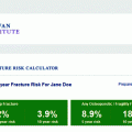
A patient with a T-score between −1.0 and −2.5 at the lumbar spine or femoral neck and who has a FRAX® 8 10-year probability of hip fracture of ³3 % or a FRAX® 10-year probability of any major osteoporotic fracture of ³20 %, using the US-adapted WHO algorithm
Treatment Guidelines from AACE and NAMS
The 2010 American Association of Clinical Endocrinologists (AACE) guidelines [21] for the treatment of postmenopausal osteoporosis are identical to those from the NOF, although AACE issued them in the context of treating postmenopausal osteoporosis only. The treatment guidelines [22] from the North American Menopause Society (NAMS) are also relevant only to postmenopausal women and, again, are identical to those from the NOF.
Treatment Guidelines from Osteoporosis Canada and NOGG
Guidelines from both Osteoporosis Canada [23] and the National Osteoporosis Guideline Group (NOGG) in the United Kingdom [24] also state that postmenopausal women with a prior fragility fracture should be considered for pharmacologic intervention. The Canadian guidelines [23] otherwise suggest utilizing a FRAX® 9 or CAROC 10-year major osteoporotic fracture risk of >20 % to identify individuals as being at high risk and candidates for pharmacologic therapy. In individuals at moderate fracture risk based on a 10-year major osteoporotic fracture risk of 10–20 %, other factors which may warrant consideration of pharmacologic intervention include a lumbar spine T-score that is much lower than the femoral neck T-score, a wrist fracture in an individual over age 65 who also has an osteoporotic BMD T-score, rapid bone loss, recurrent falls, men receiving androgen-deprivation therapy10, and women receiving aromatase-inhibitor therapy. The guidelines for pharmacologic intervention in the absence of a prior fragility fracture from NOGG are also based on FRAX® 10-year major osteoporotic fracture risk, but the intervention threshold based on that risk varies according to the patient’s age. Graphs illustrating the recommended intervention threshold based on 10-year fracture risk and age can be found in the NOGG Pocket Guide for Healthcare Professionals which is available online at http://www.shef.ac.uk/NOGG/downloads.html.
Interventions in Osteoporosis
Interventions in osteoporosis are divided into two basic categories: nonprescription and prescription. The nonprescription interventions can be further divided into lifestyle modifications and over-the-counter supplements or medications.
Nonprescription Interventions
Lifestyle Modifications
Many lifestyle modifications recommended to prevent bone loss and fractures are modifications that are appropriate for everyone in general, not just the woman concerned about osteoporosis. In some cases, however, the recommendations are different if the woman already has a very low bone density or fracture compared to the woman who has a normal bone density and is concerned about future bone loss. Recommendations that are appropriate for everyone include:
Avoidance of cigarette smoke
Moderation in alcohol intake
Moderation in caffeine intake
Moderation in salt (sodium) intake
Modification of the home environment to reduce the risk of falls
Cigarette smoke, alcohol, caffeine, and sodium are all associated with bone loss to some degree [25–29]. Modification of the home environment does not have to be extensive to reduce the risk of falls. Measures include removal of throw rugs; elimination of electrical extension cords from walk areas; installation of automatic night-lights in the bedroom, bath, and kitchen; and installation of safety bars in the bath. Only the installation of safety bars in the bath requires any expertise or potential expense. The bars themselves are not expensive, but they must be installed into the studs of the wall with long screws in order to support the weight of the body. These are relatively simple measures that can be lifesaving.
Calcium, Vitamin D, and Exercise
Nonprescription interventions that are appropriate for most, but not all, women are obtaining adequate calcium and vitamin D and regular weight-bearing or resistance exercise. The NOF recommends an elemental calcium intake from all sources combined of 1,200 mg a day [20]. For men and women aged 50 and older, the NOF recommends 800–1,000 I.U. of vitamin D per day. The recommended dietary allowance11 (RDA) for calcium according to the Institute of Medicine (IOM) of the National Academies of Science is 1,200 mg a day for women aged 51–70 and 1,000 mg a day for men of the same age. After age 70, 1,200 mg a day is recommended for both men and women [30]. The RDA for vitamin D from the IOM is lower than the amount suggested by the NOF at 600 I.U per day for both men and women aged 51–70. After age 70, the IOM recommends 800 I.U. per day. Although the recommendations for calcium intake from the two groups are similar, the IOM recommendations for vitamin D intake are more conservative than those of the NOF and are considered by some to be inappropriately low [31]. The recommended amounts of vitamin D from both groups are quite safe however. The upper limit of safe intake for vitamin D, as set by the IOM, is currently 4,000 I.U. per day [30].
Obtaining the recommended amounts of calcium from the diet alone is often difficult but preferred. Over-the-counter calcium supplements are an acceptable means of supplementing dietary calcium to ensure that the intake goals are met. Calcium supplements are relatively inexpensive. Most supplements are forms of calcium carbonate, but supplements of calcium citrate, calcium phosphate, and combinations of calcium lactate and gluconate are also available. All of these supplements with the exception of calcium citrate should be taken with food. Calcium citrate is best taken on an empty stomach. It is also important to note the mg of elemental calcium per tablet, as this is the important amount, not the mg of calcium salt. For example, a common strength of calcium carbonate tablet is 1,250 mg of calcium carbonate. This tablet size provides 500 mg, not 1,250 mg, of elemental calcium. Calcium-fortified foods and beverages are also useful in increasing dietary calcium intake. There are a variety of fruit juices that are now fortified with calcium as well as calcium-fortified bread, rice, and cereal. Obtaining excessive amounts of calcium is not recommended. There is no proof that consuming amounts in excess of those recommended is beneficial. It may also be possible to increase the likelihood of developing a kidney stone if excessively large amounts of calcium are excreted into the urine.
Patients should always be asked if they have ever had a kidney stone or been told to avoid foods high in calcium before recommending an increase in dietary calcium or calcium supplements. If they have, they should be encouraged to discuss this issue with their physician before proceeding on their own.
Vitamin D is important for calcium absorption and metabolism, but it is not necessary to have vitamin D and calcium in the same tablet. The vitamin D in over-the-counter preparations must undergo chemical conversions in the liver and kidney before becoming biologically active. Because of this delay in becoming active, any vitamin D combined with a calcium supplement does not actually affect the absorption of the calcium in that supplement. Its actions will begin later. Although vitamin D is made in the skin from the interaction of UVB rays from sunlight and a form of cholesterol found in the skin, vitamin D supplements are generally necessary to meet the recommended amounts. The amount of vitamin D made in the skin from sun exposure is affected by the geographic location of the individual, the time of year, the time of day, the length of the exposure, and the age of the individual. The use of sunscreen with an SPF greater than 8 will block up to 95 % of the production of vitamin D. Fortunately, vitamin D supplements are inexpensive and readily available without a prescription. It would seem practical to utilize a supplement rather than worry if sufficient, unprotected sun exposure had been achieved.
Exercise is important in bone health. The most beneficial forms of exercise for the skeleton are weight-bearing exercise and resistance exercise. Weight-bearing exercise is any type of exercise that forces the skeleton to support the weight of the body. For example, walking is a weight-bearing exercise, whereas swimming is not. Resistance exercise is exercise in which the muscles push or pull against a resistance. Such resistance can be in the form of weight, tension, or air pressure. This is the type of exercise that is performed with the use of machines or free weights. There are restrictions on the type of exercise that women with osteoporosis should perform. High-impact exercises may place a fragile spine at risk for fracture and should be avoided. This includes running, rope jumping, and high-impact aerobics. Any type of exercise in which the risk of falling is increased should be avoided. Exercises that require repeated or resisted trunk flexion12 should also be avoided since this may increase the risk of spine fracture. Such exercises include traditional sit-ups and toe-touches. Trunk or spine extension exercises are both safe and recommended.
Prescription Interventions
The prescription medications used in osteoporosis are either antiresorptive or anabolic agents. Antiresorptive agents are approved by the Food and Drug Administration (FDA) for the prevention of osteoporosis, for the treatment of osteoporosis, or both. An antiresorptive agent is a medication that primarily inhibits bone loss rather than stimulating new bone formation. Increases in the measured bone density are observed with these agents, which may seem contradictory if the agents primarily inhibit bone loss. Part of this increase is attributed to the agent stopping additional bone loss while the skeleton rebuilds bone naturally. The bone rebuilding is not being directly stimulated by the antiresorptive agent. The term “anti-catabolic” may be used instead of antiresorptive. This term has not been widely adopted, but its use may be encountered in clinical practice. Estrogen, raloxifene, calcitonin, bisphosphonates, and denosumab are antiresorptive or anti-catabolic agents. There is only 1 anabolic agent, teriparatide, which is currently approved by the FDA for the treatment of osteoporosis. Anabolic agents stimulate bone formation rather than inhibit bone loss.
Agents that have been approved by the FDA for the prevention of bone loss or the prevention of osteoporosis are agents that have been shown to inhibit bone loss. Agents that are FDA-approved for the treatment of osteoporosis have generally been shown in clinical trials to reduce the risk of fractures. The exception to this is in the treatment of osteoporosis in men in which the specific FDA-approved indication is for the treatment of osteoporosis in men to “increase bone mass.” Physicians are not restricted by these approvals from using a medication to treat osteoporosis that has been approved only for prevention and vice versa if it is deemed medically appropriate to do so.
Estrogen Therapy
A number of estrogen preparations are approved for the prevention of osteoporosis. The approval for the prevention of osteoporosis does not extend to every form of estrogen that is available by prescription. It is given only to those preparations that have provided information from clinical trials to the FDA, demonstrating that the particular preparation at a specific dose inhibits bone loss.
The Selective Estrogen Receptor Modulator Raloxifene
Raloxifene, or Evista®


Related posts:

Stay updated, free articles. Join our Telegram channel
Full access? Get Clinical Tree
