Anal Carcinoma
Todd M. Blodgett, MD
Alex Ryan, MD
Omar Almusa, MD
Key Facts
Terminology
Anal cancer, squamous cell carcinoma (SCCA) of the anus
Imaging Findings
Most lesions arise in anal canal
Consider PET/CT for staging
MR and endoluminal US commonly used to assist in determining depth of penetration and local spread
Post-treatment PET/CT results are more predictive of survival outcome than pre-treatment factors such as T-stage and nodal status
Patient with partial metabolic response, i.e., persistent FDG uptake in irradiated region, have 2 year progression-free survival rate of 22%
Best imaging features include focal intense FDG activity on a PET/CT scan with a correlative CT abnormality ± inguinal/iliac nodes
Focal intense FDG activity is usually identified within the primary lesion if large enough (> 6 mm)
Regional lymph nodes including inguinal, perirectal, and iliac nodes may be involved
Top Differential Diagnoses
Physiologic FDG Activity at the Anorectal Junction
Metastatic Disease
Distal Colonic Adenocarcinoma
Rectovaginal Fistula
Inflammation of the Anus
Diagnostic Checklist
FNA of inguinal adenopathy helpful to establish histologic proof of metastasis
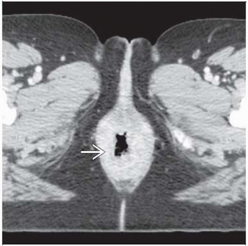 Axial CECT shows massive diffuse thickening of the anal mucosa  in this patient with recently diagnosed squamous cell carcinoma of the anus. in this patient with recently diagnosed squamous cell carcinoma of the anus. |
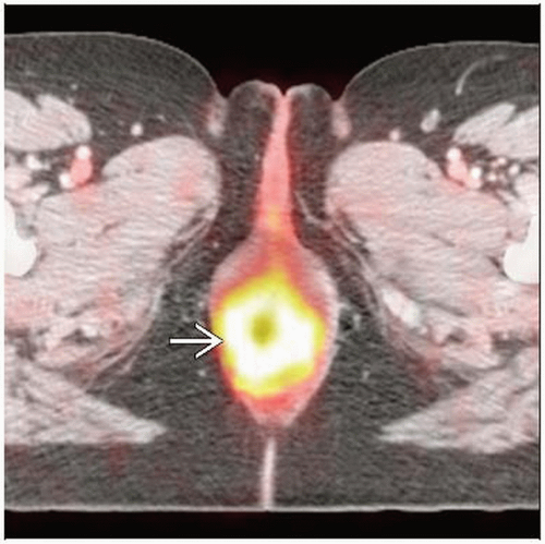 Axial fused PET/CT shows correlative circumferential intense FDG activity corresponding to the anal mucosal thickening  . . |
TERMINOLOGY
Abbreviations and Synonyms
Squamous cell carcinoma (SCCA) of the anus
Anal carcinoma
Anal cancer
Definitions
Carcinoma arising from tissue of the anal canal or anal margin
Subclassified as transitional and cloacogenic
IMAGING FINDINGS
General Features
Best diagnostic clue
Usually diagnosed by physical exam
Best imaging features include focal intense FDG activity on a PET/CT scan with a correlative CT abnormality ± inguinal/iliac nodes
Location
Most lesions arise in anal canal
Anatomic area extends from anorectal ring to zone approximately halfway between pectinate (dentate) line and the anal verge
Carcinomas arising proximal to pectinate line (transitional zone between glandular mucosa of rectum and squamous epithelium of distal anus)
Basaloid, cuboidal, or cloacogenic tumors
About 1/3 of anal cancers have this histology
Malignancies distal to pectinate line are of squamous histology
Account for 55% of all anal cancers
Ulcerate more frequently
Dentate line and extending approximately 1 cm proximally
Transitional zone of epithelium that connects squamous cell epithelium of anoderm with columnar epithelium of rectum
Transitional zone includes columnar, cuboidal, transitional, and squamous epithelial cells
Represents the source for a variety of malignancies that arise in the anal canal
WHO divides anal canal into 2 regions for grading malignancies
Anal canal portion: Proximal to dentate line and including transitional zone
Anal margin: Anoderm distal to dentate line
Anal canal malignancies metastasize to
Mesenteric lymph nodes and portal circulation
Regional inguinal nodes and via systemic circulation
Nodal dissemination pathways commonly target perirectal, iliac, and inguinal basins
Distant spread frequently involves liver and lung
Metastases to spine and musculoskeletal system are rare
Size: Variable, ranging from subcentimeter to several centimeters
Imaging Recommendations
Best imaging tool
MR and endoluminal US are commonly used to assist in determining depth of penetration and local spread
Consider PET/CT for regional and distant staging
Protocol advice
Immediate voiding prior to PET is recommended to minimize FDG activity in the bladder
Scan from bottom to top to minimize FDG accumulation in the bladder during the exam
Nuclear Medicine Findings
PET
Focal intense FDG activity is usually identified within primary lesions > 6 mm
Regional lymph nodes including inguinal, perirectal, and iliac nodes may be involved
Usually FDG avid unless small
FDG PET for prognosis
Complete metabolic response was associated with significantly improved progression-free and cause-specific survival compared with partial response
Patients with complete metabolic response had 2 year progression-free rate of 95%
Patients with partial metabolic response, i.e., persistent FDG uptake in irradiated region, have 2 year progression-free survival rate of 22%, regardless of presenting T-stage
Post-treatment PET/CT results were more predictive of survival outcome than pre-treatment factors such as T-stage and nodal status
DIFFERENTIAL DIAGNOSIS
Physiologic FDG Activity at the Anorectal Junction
Very common and the most likely alternative diagnosis
No correlative CT abnormality
Metastatic Disease
Rare
Occasionally seen with melanoma or local metastases from cervical, ovarian, or other pelvic malignancies
Distal Colonic Adenocarcinoma
May involve the anus
Rectovaginal Fistula
Associated with pelvic irradiation
Inflammation of the Anus
Indistinguishable from small malignancy or physiologic activity on PET and PET/CT
PATHOLOGY
General Features
General path comments
Squamous cancers make up majority of anal malignancies
Anal margin neoplasms include Bowen disease, squamous cell carcinoma, basal cell carcinoma, and Paget diseaseRelated posts:

Stay updated, free articles. Join our Telegram channel

Full access? Get Clinical Tree


