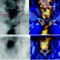
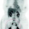
Fig. 62.1
The MIP image shows metabolic response of the left lung mass but there is a clear progression of disease in all mediastinal and abdominal injuries. Moreover, we find the appearance of new metastatic lymph nodes in the left clavicular fossa and in the ipsilateral region 81 lateral cervical

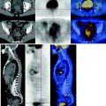
Fig. 62.2
The coronal reconstruction of CT-PET performed before radiation therapy shows the metabolism of the left lung neoplasm, presenting with dendritic branches in connection with the parietal pleura and mediastinum

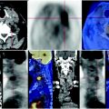
Fig. 62.3


Before the radio-chemotherapy, CT-PET shows the dorsal segment of the upper lobe of the left lung in close contact with the posterior parietal pleura, a solid speculated mass with irregular margins, which has extensive metabolism. Lymphadenopathy is associated with increased deposition of FDG at the level of the aorto-pulmonary window and right hilum
Stay updated, free articles. Join our Telegram channel

Full access? Get Clinical Tree


