OF THE FETAL
HEART
 FETAL VISCERAL SITUS
The first step in the ultrasonographic evaluation of the fetal heart is the assessment of the fetal visceral situs (laterality of fetal organs). Establishing the fetal visceral situs allows for an accurate determination of ventricular and atrial situs and therefore should be part of every fetal ultrasound examination. Three types of visceral situs exist: situs solitus, situs inversus, and situs ambiguous (Table 4-1). Situs solitus refers to the normal arrangement of vessels and organs within the body. Situs inversus, with an incidence of about 0.01% of the population, refers to a mirror-image arrangement of organs and vessels to situs solitus. Situs inversus is associated with a slight increase in the incidence of complex congenital heart disease (CHD), in the order of 0.3% to 5% (1). Furthermore, in about 20% of cases of situs inversus, Kartagener syndrome is noted, which primarily involves ciliary dysfunction with recurrent respiratory infection and reduced fertility (2). Situs ambiguous (heterotaxy), which refers to visceral malpositions and malformations different from situs solitus or inversus, is commonly associated with complex congenital heart disease, abnormalities in venous drainage, bowel malrotations and obstruction, and splenic, biliary, and bronchial tree abnormalities. The incidence of situs ambiguous has been estimated around 1 per 10,000 infants (3). Two types of heterotaxy exist: right isomerism and left isomerism. In right isomerism, also referred to as asplenia, both sides of the body show the right morphology; in left isomerism, also referred to as polysplenia, both sides of the body show the left morphology. Chapter 22 presents a detailed overview of fetal heterotaxy.
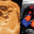
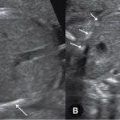
Although the current method for the determination of fetal situs relies on the position of the stomach and heart in the abdomen and chest, respectively, careful attention should be paid to the position of the aorta and inferior vena cava below the diaphragm, the presence of bowel dilatation, the presence of a gallbladder, and the presence and location of the spleen (Fig. 4-1). It is generally agreed that the positions of the aorta and inferior vena cava below the diaphragm are more reliable criteria for the determination of right or left isomerism.
Related posts:

Stay updated, free articles. Join our Telegram channel

Full access? Get Clinical Tree


