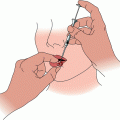
Fig. 1
Patient with squamous cell carcinoma of the skin with clinical perineural invasion of the second division of the trigeminal nerve and the facial nerve. Gross tumor extends through the skull base and invades the left cavernous sinus (arrow)
Patients with PNI have an increased risk of metastases to the regional lymph nodes [4, 9, 27] which can also be assessed on MRI but preferably with contrast-enhanced CT. Sentinel lymph node biopsy (SLNB) may also be considered to assess the regional nodes [28].
Treatment
Patients with microscopic PNI have almost always undergone excision of the primary lesion prior to the diagnosis. Whether or not to routinely add postoperative RT following complete excision is controversial. A survey of approximately 25 % of the membership of the American College of Mohs Surgery revealed that most respondents considered in-transit metastases and PNI as the major factors leading to consideration of radiographic staging, SLNB, and adjuvant RT for patients with high-risk SCC [15]. In 2009, Jambusaria-Pahlajani et al. [29] reported on a literature review of reports on high-risk SCC with PNI treated with either surgery alone or surgery and adjuvant RT. They observed that, for 74 patients with PNI, the outcomes after surgery alone and surgery plus RT were similar and concluded that, in the presence of clear surgical margins, the benefit of adjuvant RT was unclear. The disadvantage of retrospective literature reviews is that the patients at higher risk for recurrence are probably more likely to receive adjuvant RT, thus biasing the comparison. In relation to the surgical technique, Mohs micrographic surgery (MMS) is likely superior in terms of local control to standard surgical resection in SCC with PNI.
In 2009, Ross et al. [12] reported that the diameter of the nerve involved with PNI may be useful to predict outcome with PNI. They reported on 48 patients with SCC and PNI treated at the University of Pennsylvania between 1996 and 2005 with surgery (46 patients), RT alone (1 patient), and desiccation and curettage (1 patient) [12]. Surgery was combined with adjuvant RT in 25 patients. The median diameter of involved nerves was 0.09 mm (range, 0.02–0.6 mm). Overall, 17 % of patients died from recurrent SCC. PNI of nerves less than 0.1 mm was associated with a cause-specific death rate of 0 % compared with 32 % for those PNI of larger caliber nerves. Other parameters significantly associated with diminished survival included recurrent or poorly differentiated SCC, tumor diameter ≥2 cm, and/or depth of invasion of ≥1 cm. Thus the diameter of the involved nerve may be useful to select patients for treatment with surgery alone.
The indications for postoperative RT in patients with incidental PNI are not well-defined. A subset of patients with focal incidental PNI with negative margins is likely to be cured with surgery alone, particularly a BCC that is not adjacent to a major CN. Alternatively, those with multifocal PNI should be considered for postoperative RT, particularly patients with recurrent cancers, SCCs, and mid-face locations in proximity to CNs V and VII. Patients with extensive microscopic PNI associated with BCC have a high risk of recurrence with extension to the skull base after surgery alone and should be considered for postoperative RT. Patients who are immunocompromised due to solid organ transplant and/or chronic lymphocytic leukemia tend to have more aggressive cancers and should be strongly considered for postoperative RT.
In practice, the majority of patients with SCC and microscopic PNI undergo postoperative RT to reduce the risk of locoregional recurrence. RT fields usually encompass the primary site with a surrounding margin. With extensive PNI with positive margins, the involved nerve is included in the RT treatment volume to the skull base. The latter patients experience more treatment-related toxicity because of the increased volume of tissue irradiated and have a worse prognosis due to selection bias [23]. The risk of lymph node metastases is approximately 15–20 % in patients with SCC and microscopic PNI, hence clinically negative regional nodes should be electively treated [23, 24]. Although SLNB may be considered, our practice is to forego the procedure.
Patients with clinical PNI have a worse prognosis and their management is less controversial. If the gross disease appears to be completely resectable, they are treated with resection and postoperative RT. A significant subset of patients with tumor that extends proximally to the skull base has unresectable disease. Aggressive subtotal resection of such tumors, such as those that involve the cavernous sinus, results in multiple CN deficits and significant residual disease without meaningfully enhancing the probability of cure. Thus, such patients are better treated with definitive RT. Treatment volumes are individualized based on the location and extent of the tumor. The volume is initially generous because the extent of subclinical disease is often difficult to define. The clinician must be aware that the tumor involving CN V may involve CN VII via the auriculotemporal nerve, or vice versa [30]. Patients are treated with hyperfractionation at 1.2 Gy per fraction twice daily to doses in the range of 64.8–74.4 Gy postoperatively (depending on the margins) and 74.4 Gy for definitive RT. Hyperfractionation is preferred to once-daily fractionation to reduce the risk of radiation retinopathy and/or optic neuropathy in cases where the retina, optic nerve(s), and/or chiasm is included in the RT volume to high dose [31]. There are no data to support the use of adjuvant chemotherapy in patients with skin cancer and PNI; nevertheless, local failure is the dominant mode of recurrence in patients who are treated for clinical PNI. It may well be reasonable to extrapolate data from patients treated for mucosal head and neck SCCs and consider adding concomitant chemotherapy, such as weekly cisplatin 30 mg/m2, to improve the likelihood of local control [32].
Outcomes
In 2009, Jackson et al. [14] reported on 118 patients with cutaneous BCC or SCC and incidental (97 patients) or clinical (21 patients) PNI treated with RT between 1992 and 2000. RT was definitive in 4 patients and administered postoperatively in 114 patients. Median follow-up was 84 months (range, 4–201 months). The 5-year outcomes after treatment for incidental vs. clinical PNI were: local control, 90 and 57 % (p < 0.0001); relapse-free survival, 76 and 46 % (p = 0.003); cause-specific survival, 90 and 76 % (p = 0.002); and overall survival, 69 and 57 % (p = 0.03), respectively. Patients with microscopic PNI and BCC had improved local control compared with those who had incidental PNI and SCC, 97 % vs. 84 % (p = 0.02).
In 2011, Balamucki et al. [33] reported on 216 patients treated with curative intent at the University of Florida between 1965 and 2007 for skin carcinomas with incidental (107 patients) or clinical (109 patients) PNI. Median follow-up for living patients was 6.6 years (range, 0.6–23 years); 14 patients were lost to follow-up at a median of 35 months (range, 7.8–168.1 months). One hundred and five patients (78 %) had SCCs; the remainder had BCCs or metatypical BCCs. One hundred and thirty-three patients (62 %) had cancers that were recurrent after prior surgery.
Treatment for the 107 patients with microscopic PNI included surgery and postoperative RT (99 patients), preoperative RT and surgery (4 patients), and RT alone (4 patients) [33]. Twenty six of 107 patients (24 %) presented with clinically positive nodes. Margins were positive in 41 (40 %) of 103 patients treated surgically. The 5-year outcomes are depicted in Table 1 [33]. Seventeen of 107 patients (16 %) experienced 24 RT-severe related complications; 10 patients (9 %) required surgical interventions (Table 2). There were no fatal treatment complications.
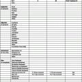
Table 1
Five-year outcomes
Five-year outcomes | Incidental PNI (N = 107) (%) | Clinical PNI (N = 109; %) | p Value |
|---|---|---|---|
Overall survival | 55 | 54 | 0.8252 |
Cause-specific survival | 73 | 64 | 0.0856 |
Local control | 80 | 54 | 0.0038 |
Local-regional control | 70 | 51 | 0.0648 |
Freedom from distant metastases | 90 | 94 | 0.1918 |
Table 2
Complications (grade ≥3)
Complication | Incidental PNI (%) N = 107 | Clinical PNI (%) N = 109 |
|---|---|---|
Soft-tissue necrosis | 5 (5 %) | 6 (6 %) |
Bone exposure | 7 (7 %) | 12 (11 %) |
Osteoradionecrosis | 3 (3 %) | 3 (3 %) |
Fistula formation | 0 | 5 (5 %) |
Wound infection | 3 (3 %) | 6 (6 %) |
Dehiscence | 1 (1 %) | 1 (1 %) |
Blindnessa | 0 | 12 (11 %) |
Other | 5 (5 %) | 9 (8 %) |
Total events | 24 | 54 |
PNI perineural invasion
Source


Related posts:

Stay updated, free articles. Join our Telegram channel
Full access? Get Clinical Tree
