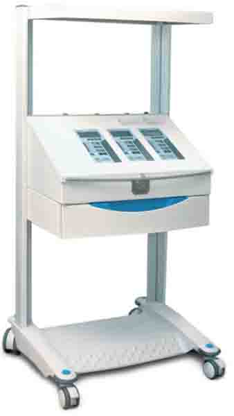
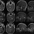
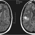
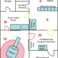
5 Anesthesia services have been provided for years to patients having magnetic resonance imaging (MRI). Caring for these patients outside of the operating room in the MRI environment is a significant challenge to any anesthesia provider. Anesthesia services are usually required for patients that cannot cooperate during the process of obtaining MRI scans. Claustrophobia, an altered sensorium, or the inability to remain still and cooperative are common indications for the involvement of anesthesia personnel in the care of these patients. MRI is an integral part of the various neuronavigational systems that are employed to perform neurosurgery in the modern era. Advances in the fields of neurosurgery and radiology combined with the development of MRI-compatible equipment have led to the development of intraoperative MRI (iMRI)-guided surgery. During an iMRI-guided procedure, the patient is imaged several times with MRI while undergoing surgery. The neurosurgeon benefits by having the ability to view the surgical site on MRI with the cranium open before the conclusion of the procedure. Although the provision of anesthesia for iMRI is similar to that during standard MRI, additional complexities related to the surgical aspect of the procedure exist. During iMRI, MRI is an essential part of the operation. The iMRI provides the surgeon with an intraoperative way to monitor the extent of the tumor resection as well as the ability to compensate for brain shift during microscope-based neuronavigation.1,2 This MRI environment is clearly different than that encountered with conventional MRI and the anesthetic care of the patient must be adapted to this new circumstance and its unique complexities. For the past decade, neurosurgeons have used various neuronavigation systems to perform precise and skillful surgical procedures. Surgical applications for iMRI include transsphenoidal resection of pituitary tumors, deep brain stimulation electrode placement for Parkinson’s disease,1 cerebral abscess drainage,2 tumor removal,3 and brain biopsy.4 iMRI allows near-real time three-dimensional imaging of the brain once the cranium is open, unlike static standard MRI of the brain.5 There is little doubt that the anesthesiologist will become more involved in the care of patients undergoing neurosurgical procedures during an iMRI-guided surgical procedure where the imaging is a crucial part of the surgery. Many anesthesiologists are uncomfortable providing anesthesia in MRI suites because of the technical issues associated with the environment. With a better understanding of the numerous challenges facing the anesthesia team, an effective plan can be formulated and implemented that will minimize patient risk and reduce procedural morbidity and mortality. The goal of the anesthesiologist should be to provide a safe anesthetic for the patient and further contribute to the completion of surgery in an organized and efficient manner. Although significant challenges and obstacles are present during iMRI-guided surgery, anesthesia care for these patients can be safe and effective by adhering to strict standards. MRI uses high-powered magnetic fields and radiofrequency (RF) pulses to produce digitalized topographic images. Because MRI does not employ ionizing radiation, there are no known physiologic effects experienced by the patient. Difficulties in providing anesthesia services in the MRI suite result primarily from the magnetic field strength and the narrow cylindrical imaging space of most high-field (≥ 1.5 tesla) MRI scanners. In general, it is understood and accepted that patients must be closely monitored under anesthesia and during surgery. The guidelines published by the American Society of Anesthesiology for nonoperating room locations where anesthesia is provided were first approved in October of 1994 and last amended in 2003.6 During MRI and iMRI-guided surgery, physiologic monitors must function properly within the strong magnetic field. For a variety of reasons, standard anesthesia machines cannot be used during MRI. These reasons include changing magnetic fields and RF currents, which will cause interference and/or malfunction of the monitoring equipment. Additionally, there is electrical interference between the physiologic monitoring equipment and the MRI scanner and vice versa. Not only do the monitors function poorly in a magnetic field, the images obtained during MRI can be distorted by the electronics inherent to the monitoring equipment. Any object made of metal that is ferromagnetic can be attracted by the strong magnetic fields. This attraction can cause physical injury to personnel in the room and can result in equipment damage. Most MRI suites have a test magnet for the express purpose of identifying ferromagnetic equipment prior to it entering the room. Non-ferromagnetic implants are less problematic, but are not completely without their unique risks. The intermittent RF pulses used during MRI can cause heat generation within a metal object. Conducting wires can arc under certain favorable conditions such as when they are conducting electrical current. 1. Patient accessibility and their distance from the anesthesia source 2. The need to move the patient into the MRI scanner 3. The length of the operative procedure 4. The availability of trained personnel to provide assistance during transport from the operating room and postanesthesia care unit 5. The risk of thermal injury and RF interference 6. The migration of ferromagnetic instruments 7. Inability of anesthesia providers to hear acoustic monitor alarms during the scanning process In general, the goal of the anesthesiologist providing anesthesia during MRI is to provide a safe anesthetic to a quiet, comfortable, and immobile patient. For standard MRI, sedation may be all that is necessary in contrast to the majority of iMRI cases where a general anesthetic is more appropriate. Patient movement during imaging can severely degrade the MR image quality. Only during specific neurosurgical procedures such as an awake craniotomy, should a patient receive sedation alone as their anesthetic. The preoperative anesthesia evaluation for iMRI-guided surgery is similar to the assessment for any neurosurgical patient. The strong magnetic fields used during iMRI-guided surgery can dislodge ferromagnetic objects including those implanted in patients. These include cerebral aneurysm clips as well as metallic intraocular foreign bodies. Safe metals that are not ferromagnetic include titanium,7 silver, gold, and aluminum. Special attention should be paid to patients with pacemakers (including automatic implantable defibrillators-pacers) and implanted infusion pumps. Pacers and automatic implanted defibrillators will become inactivated in a magnetic field.8 Patients with these implanted devices are not good candidates for an iMRI-guided surgical procedure. In procedures where the patient may be awake or receive monitored anesthesia care, contraindications include those conditions indicated above in addition to patient factors that may influence the overall optimal MR image quality. Factors such as the psychologic make-up of the patient can preclude patient selection for awake procedures using iMRI-guidance. Those patients with anxiety disorders, the inability to be cooperative, a fear of closed spaces or claustrophobia, and the inability to understand or comprehend the expectations that are required of them during the procedure may negate their candidacy for this type of surgery. Every effort must be made to use MRI-compatible equipment when performing iMRI-guided surgery. MRI-compatible equipment has filtered electrical current that does not interfere with the performance of the magnet. Equipment used in an MRI environment must meet the following criteria: 1. It has to function properly. 2. It should pose no danger to patient, personnel, and other equipment in the room. 3. It must allow the MRI scanner to obtain clear and nondistorted images. The equipment needs for iMRI-guided procedures are the same as that for conventional MRI. However, additional pieces of equipment and monitors may be needed during iMRI-guided surgery. Because of the need for high magnetic field strength and the infrastructure necessary to create these fields, the iMRI suite is usually separate from the main operating room. Sufficient work space must be available for anesthesia personnel in the iMRI-guided surgery suite to function without being handicapped. Rooms adjacent to the iMRI surgical suite may be necessary for storage of anesthesia equipment and of other utilities. The anesthesia provider should have an adequate supply of drugs, infusion pumps (Fig. 5.1), and other equipment with common spare parts to perform a safe anesthetic in the iMRI suite. Common items needed for anesthesia care should be routinely stocked separately and in proximity to the iMRI suite. These items should include a large selection of drugs, intravenous medications, and pumps, as well as airway management equipment in the event that a difficult airway is encountered. Nonanesthesia personnel working at these remote locations are usually not familiar with the unique requirements of providing anesthesia and can be of little assistance if there is an anesthesia-related emergency. In the past, some have advocated having all equipment outside of the magnetic field. The gauss line or the point where ferromagnetic items are attracted to the magnet is between 30 to 50 gauss lines.9 Image quality is proportional to the magnetic field strength of the scanner. The unit used for quantifying the magnetic field strength is a tesla (T) or 10,000 gauss. Scanners used for iMRI-guided surgery are between 0.12 T and 3 T. To be entirely outside of the magnetic field, the ventilator and monitoring equipment would need to be up to 20 feet away from the scanner. Very long ventilator breathing circuits, intravenous lines, and pressurized monitoring tubing would be necessary.10 Fig. 5.1 Tesla infusion pumps. (Courtesy of Mammendorfer Institut für Physik und Medizin GmbH.) Monitors that use batteries would need them to be safely secured because they are highly ferromagnetic. To avoid electrical interference from monitoring equipment, it is best to have the iMRI suite powered by filtered electrical current.
Anesthesia Considerations
Issues Associated with MRI and Anesthesia Administration
Other Obstacles to Providing Optimal Anesthetic Care in an MRI Environment
Contraindications to iMRI
Anesthesia Equipment
Anesthesia Machine
Related posts:
Stay updated, free articles. Join our Telegram channel

Full access? Get Clinical Tree


