Aneurysmal Subarachnoid Hemorrhage
Kwaku A. Obeng
CLINICAL HISTORY
56-year-old female presenting with acute onset of a severe headache.
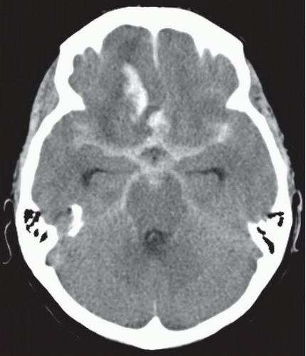 FIGURE 61A |
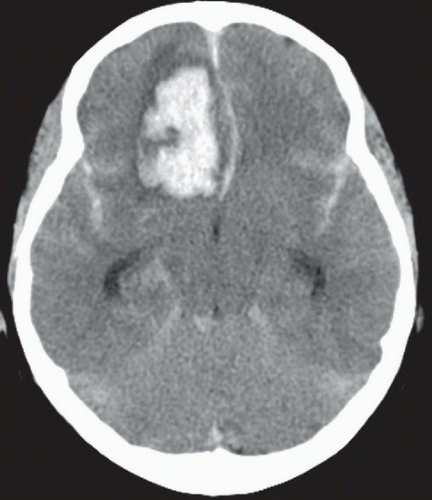 FIGURE 61B |
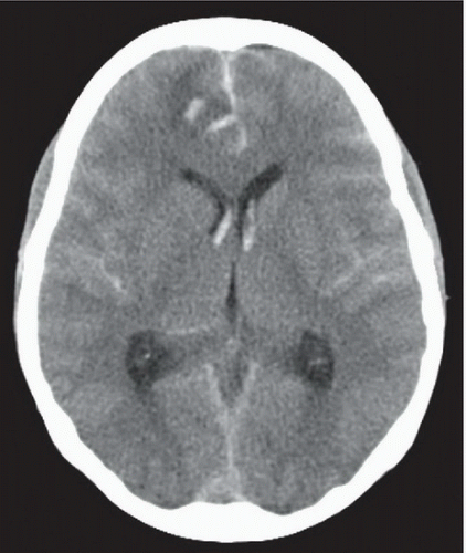 FIGURE 61C |
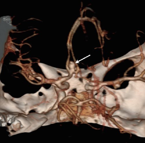 FIGURE 61D |
FINDINGS
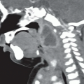
Figures 61A,61B and 61C: Axial noncontrast head CT images from inferior to superior demonstrate diffuse subarachnoid hemorrhage in the suprasellar, interpeduncular, and ambient cisterns with extension laterally into the Sylvian fissures and superiorly along the interhemispheric fissure. There is also a parenchymal hematoma in the right frontal lobe (most apparent in Figs. 61A and 61B) as well as hemorrhage in both lateral ventricles (Fig. 61C). Figure 61D: 3D surface shaded reconstruction from a CTA of the circle of Willis demonstrates a superiorly oriented saccular aneurysm (arrow) arising from the anterior communicating artery.
Related posts:


Stay updated, free articles. Join our Telegram channel

Full access? Get Clinical Tree


