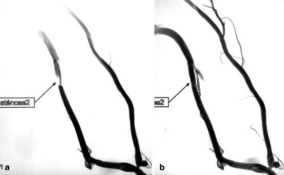
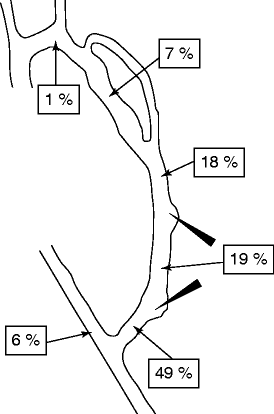
Fig. 9.1
(a, b) This angiogram of a left radial–cephalic AVF referred for low flow shows an apparently normal proximal radial artery but abnormally well-developed collaterals running from the interosseous artery to the distal radial artery, whose flow is reversed. These collaterals, resembling an arteriovenous malformation, are especially developed because the palmar arch and the distal radial artery are stenosed at the wrist and cannot compensate for the insufficient inflow of the proximal radial artery. Such a development of interosseous collaterals is pathognomonic of a stenosis of the proximal radial artery, which was not visible on the anteroposterior runs. (c) The severe anastomotic stenosis of the proximal radial artery becomes visible only on the first frame of this lateral run and is then subsequently masked by the superimposition of the opacification of the vein
As a general rule, the proximal radial artery feeding a forearm AVF should be of larger caliber and opacify earlier than the ulnar artery. If this is not the case, then the radial artery is likely diseased and may not sufficiently expand to provide enough flow to the AVF given that the diameter of the artery determines its volume flow. On the other hand, the distal radial artery should flow retrogradely, that is, toward the anastomosis rather than toward the hand. Antegrade distal radial artery flow is suggestive of low access flow caused by tight stenosis of the fistula. A distal radial artery larger in size than the proximal segment invokes a proximal artery stenosis or diffuse artherosclerotic infiltration.
In long-standing upper arm AVFs, the entire proximal brachial artery flow may be directed into the arterialized vein with no opacification of the brachial artery distal to the anastomosis (Fig. 11.1a, b). Flow in distal arterial segment is maintained by collaterals arising from the more proximal axillary or proximal brachial artery segments.
The diagnosis of a localized short-segment arterial stenosis is usually straightforward. Congenital arterial lesions like fibrodysplasia are not rare, but stenosis grading at their level may be challenging.
9.5.3 The Veins
The arterialized vein caliber should be larger than its feeding artery unless if it is stenosed. Not all stenoses are detrimental and therefore justify dilation. For instance, a juxta-anastomotic stenosis in a hyper flow AVF serves to protect the fistula from high pressure. Flow into collateral or accessory veins indicates the presence of a downstream critical stenosis of the main vein but can also be a sign of hyper flow whereby all recruitable veins are opacified. Collaterals should be clearly distinguished from normal anatomy. The main cephalic vein and accessory cephalic vein usually opacify in the forearm, while the basilic and cephalic vein in the upper arm. However, reflux of contrast all the way to the wrist is abnormal.
Isolated drainage into the perforating vein at the elbow suggests occluded cephalic and basilic veins. In the eyes of a beginner, a deep brachial vein can easily be mistaken for the basilic vein. They can be differentiated by taking an orthogonal view of the elbow or upper arm. It is frequently possible to identify the stump of an occluded basilic or cephalic vein that may be recanalized. Reopening such a direct superficial drainage at the elbow is critical to increasing long-term survival of forearm AVFs.
Venous spasm can easily be mistaken for stenosis. Spasms are induced by endovascular devices like introducer sheaths and guidewires. Pseudo-stenosis can also arise at the point of injection of local anesthetic, which produces an occlusive mass effect, particularly on the vein lumen. It is best to repeat the angiography with a tourniquet at the end of the intervention to ascertain whether it has disappeared.
A stenosis is defined as a narrowing of the vessel lumen referenced to an adjacent normal upstream and downstream vessel segment. The reference vessel is very arbitrarily and subjectively defined and can pose challenges when infiltrated or aneurysmal. Hence, to be certain of the presence of a stenosis, it is always best to take two orthogonal views of the narrow vessel segment in question—frames taken with forearm supinated and pronated in the case of distal AVFs. Such techniques also allow proper detailed viewing and studying of the forearm to rule out any entrapment syndromes of the radial or ulnar arteries [8].
Stenoses can be masked by superimposed adjacent aneurysmal segments (Fig. 9.1). Hypertrophic and sometimes calcified valves can be the cause of a very focal stenosis, and this can be easily missed on angiography except on the initial frames where it appears as a narrow jet of contrast (Fig. 9.2a, b), which then rapidly blends in with the valves and oncoming rush of contrast.


Fig. 9.2
(a) This basilic vein stenosis due to hypertrophic valves is visible only in the form of a jet of contrast on the first frame. (b) It is subsequently overwhelmed by the full venous opacification on later frames
Stay updated, free articles. Join our Telegram channel

Full access? Get Clinical Tree


