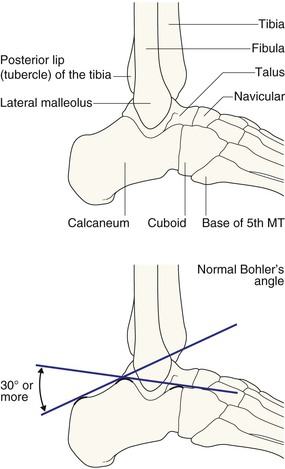
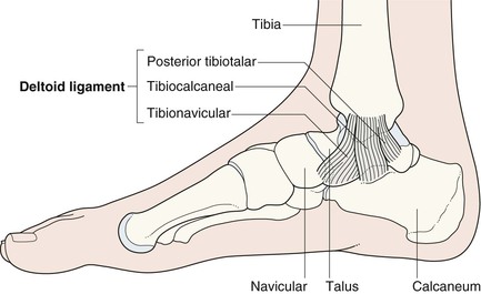
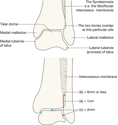
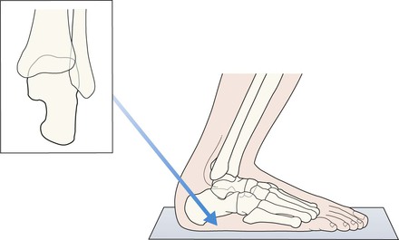
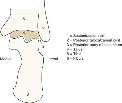
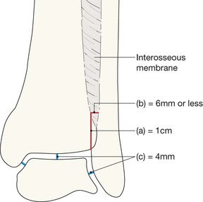
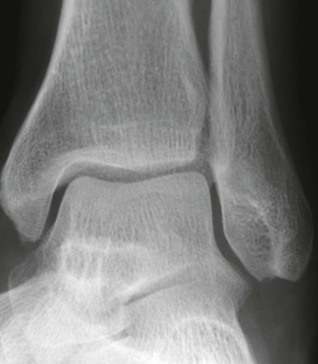
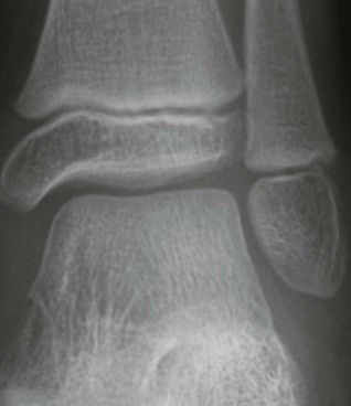
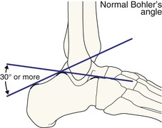
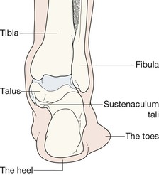
The lateral and medial malleoli can be identified. Helpful hints to aid identification: The posterior lip (or tubercle) of the tibia, conventionally and inaccurately referred to as the posterior malleolus, is well shown. The calcaneum and its sustentaculum tali are demonstrated. Bohler’s angle can be assessed for normality. The base of the 5th metatarsal is often included. Principal ligaments—medial. The mortice projection is obtained with slight (20°) internal rotation so that the fibula does not overlap the talus. The joint space should be of uniform width all the way around. This space is well seen medially, it continues over the superior aspect of the dome of the talus, on to the lateral side of the joint. The width of the joint space measures approximately 4 mm2. The surface of the talar dome should be smooth, smooth, smooth. No irregularity, no notching, no defect. The lateral process (also known as the lateral tubercle) of the talus is an important structure. The talocalcaneal ligament attaches to this part of the bone. A useful rule: the bones of the tibia and fibula should always overlap on the mortice view. Any clear separation between these two bones should lead you to question whether the interosseous membrane is torn. The posterior two thirds of the bone is well shown; the anteriorly positioned sustentaculum tali is often not as well visualised on the radiograph. Check the: ▪ Malleoli—fracture or … fractures? ▪ Tibiofibular interosseous membrane—any suggestion of a rupture? □ If normal, the tibia and fibula should show some degree of overlap. □ A measurement: the width of the space between the distal tibia and fibula at a point 1.0 cm proximal to the tibial articular surface should not exceed 6 mm5. ▪ Talus □ Medial and lateral processes (ie the tubercles, p. 268)—any fragmentation? ▪ Joint width—does any part exceed the normal 4 mm3? ▪ Epiphyses and growth plates in children—normal? (see pp. 15 and 280). Normal features and measurements (ie rules of thumb) relating to: (1) the interosseous membrane, and (2) the various articulations at the mortice joint. Normal AP mortice view. All of the checkpoints are normal. Note that part of the fibula overlaps part of the tibia. This is a characteristically normal appearance on a mortice view. Normal AP mortice view. A child. The growth plates and all of the other checkpoints are normal. Check the: Measuring Bohler’s angle. Draw a line from the highest point anteriorly to the highest midpoint. Then draw a line from the highest point posteriorly to the highest midpoint. The angle subtended should be 30° or more. Check the: The normal standing foot seen from behind. This drawing may help you to understand the anatomy on the axial view of the calcaneum. The sustentaculum tali is the shelf-like part of the calcaneum which extends medially to support part of the head of the talus.
Ankle & hindfoot
Normal anatomy
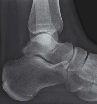
Lateral view—bones and joints
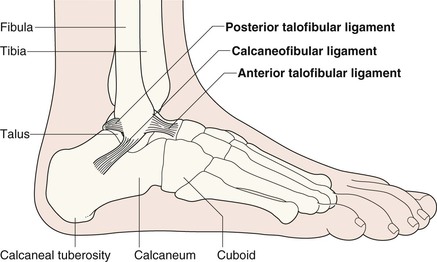
Lateral view—ligaments
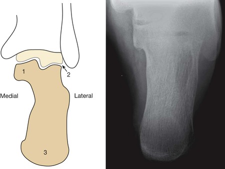
AP mortice
Axial projection—calcaneum
Analysis: the checklists1,3,4
AP mortice
Lateral view
Axial view

Ankle & hindfoot
16