(1)
Department of Radiology-Neuroradiology, “San Paolo” Hospital, Bari, Italy
A 37-year-old patient
A family history of SpA
Dorsal and lumbar pain during the night for more than 2 years
Morning stiffness and limitation of motion of the spine in the frontal and lateral planes
Limitation of chest expansion

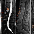
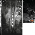
Fig. 1
X-ray of thoracic and lumbar spine. Lateral thoracic X-ray shows marginal anterior osteophytosis in the upper spine (a); lateral lumbar X-ray shows erosion of the anterior corner of L4 with a marginal osteophytosis and anterior syndesmophytosis L1–L2 (b)
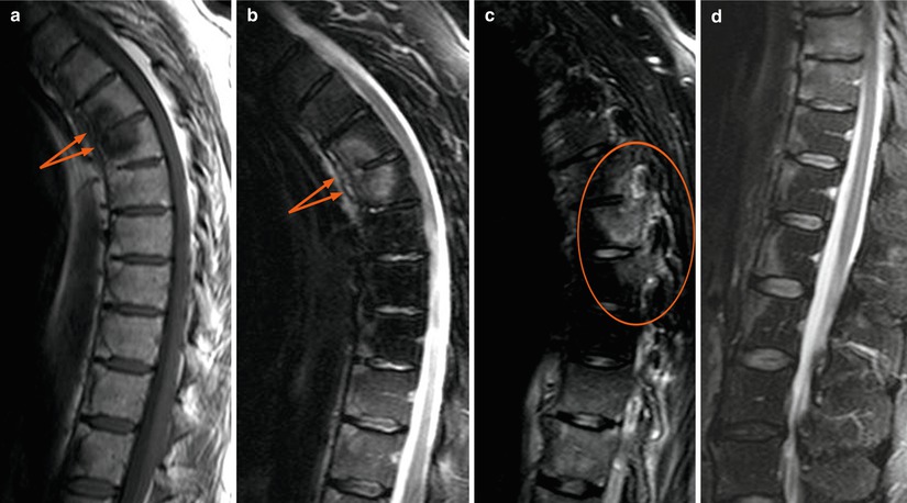
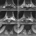
Fig. 2
Sagittal TSE T1-weighted image (a) and sagittal TSE T2-weighted images with fat saturation (b–d). The images show low signal of the T5–T6 (a, arrows) endplates and a focal fat infiltration of the vertebral corners of the thoracic spine, with ankylosis of T6–T7 and T8–T9 (a), increased signal of T5–T6 endplates (b, arrows


Related posts:
Stay updated, free articles. Join our Telegram channel
Full access? Get Clinical Tree
