The focus of this article is to illustrate various pathologic entities and variants, heralding disease about the ankle, based on scrutiny of AP radiographs of the ankle, with correlative findings on cross-sectional imaging. Many of these entities can only be detected on the AP ankle radiograph and, if not recognized, may lead to delayed diagnosis and persistent morbidity to the patient. However, a vigilant radiologist, equipped with the knowledge of the characteristic appearance and typical locations of the imaging findings, should be able to make the crucial initial diagnosis and surmise additional findings to be confirmed on cross-sectional imaging.
Key points
- •
Several characteristic locations should be scrutinized on the AP radiograph of the ankle so as to detect various disease entities which may otherwise remain undetected.
- •
Small ossific densities, protuberances, or abnormal interfaces on the AP radiograph of the ankle can be the harbingers of more extensive underlying soft tissue injury, better characterized on cross-sectional imaging.
- •
Given the clinical overlap of many of the entities described in this article with lateral ankle sprains, they may be clinically misdiagnosed and thus, the radiologist can play a crucial role by helping avoid delayed diagnosis and patient morbidity.
Introduction
Radiographic evaluation of the ankle is exceedingly common, fast, and relatively inexpensive. Often obtained to evaluate for fractures in the setting of trauma, ankle radiographs can reveal several other types of traumatic and nontraumatic lesions that may overlap clinically with lateral ankle sprain or may be unsuspected by the referring clinician at the time of injury.
The purpose of this article is to illustrate several pathologic entities and variants heralding disease about the ankle that can be initially diagnosed on anteroposterior (AP) radiograph of the ankle alone, with correlative findings on MR imaging. Many of these entities can be detected only on the AP ankle radiograph, particularly if foot films are not available, and if not recognized may lead to delayed diagnosis and persistent morbidity to the patient. However, the vigilant radiologist, equipped with the knowledge of the characteristic appearance and typical locations of the imaging findings, will often be able to make the crucial initial diagnosis and surmise additional pathologic conditions, which, if necessary, can be confirmed on cross-sectional imaging.
The article is subdivided into 2 major sections: lateral entities and medial entities. Lateral entities include extra-articular lateral hindfoot impingement, superior peroneal retinaculum (SPR) avulsion, fracture of the lateral process of the talus, fracture of the anterior process of the calcaneus, enlarged peroneal tubercle (PT), with associated peroneal tendon disease, and peroneus longus tendon tear, associated with os peroneum retraction. Medial entities include deltoid ligament injury, talar body fractures, and extra-articular (posteromedial) talocalcaneal coalition. In addition to emphasis on key radiographic and cross-sectional imaging features for each topic, the relevant anatomy, mechanism of injury, clinical features, and management are also briefly discussed.
Lateral entities
Extra-articular Lateral Hindfoot Impingement
Severe flatfoot and hindfoot valgus deformity can predispose to extra-articular, lateral hindfoot impingement, composed of talocalcaneal and subfibular impingements. With progressive hindfoot valgus alignment, the weight-bearing forces are shifted laterally from the talar dome to the lateral talus and fibula and talocalcaneal joint subluxation may develop. Despite the pathologic condition being medial, typically in the setting of posterior tibial tendon dysfunction, patients can present with lateral ankle pain because of impingement between the lateral talus and calcaneus (talocalcaneal impingement) and, as the valgus deformity progresses, between the lateral calcaneus and the distal fibula (subfibular impingement). Secondary lateral soft tissue entrapment can further contribute to pain.
On a weight-bearing AP view of the ankle, the presence of hindfoot valgus and lateral extra-articular impingement can be surmised when there is lateral tilting of the normal calcaneal axis ( Fig. 1 ) relative to the tibial axis. As impingement develops, there will be greater proximity of the fibular tip to the calcaneus ( Fig. 2 ), eventual direct contact and even a pseudofacet between the 2 bones. Imaging findings of extra-articular impingement may be delineated in greater detail with computed tomography (CT) and MR imaging, where there is compromise of the sinus tarsi space, subluxation at the subtalar joint, and apposing subcortical sclerosis, cyst formation and, in the case of MR imaging, marrow edema, at the sites of contact between the lateral talus and calcaneus and between the fibular tip and lateral calcaneus. A fibulocalcaneal pseudofacet may also be noted. MR imaging may further depict soft tissue abnormalities such as entrapment and thickening of the calcaneofibular ligament and peroneal tendon subluxation in the later stages of the disease (see Figs. 1 and 2 ).


Management of hindfoot deformity usually begins with conservative measures such as physical therapy and orthotics. Advanced osseous deformity, with the development of extra-articular impingement, typically requires calcaneal osteotomy or arthrodesis in the most severe cases.
Superior Peroneal Retinaculum Avulsion
The SPR is a band of fibrous tissue that acts as the primary restraint to peroneal tendon subluxation out of the retromalleolar groove of the distal fibula. The retinaculum originates from the posterolateral aspect of the fibula, spanning across the superficial margin of the peroneal tendons, and most commonly inserting onto the calcaneus and Achilles tendon sheath.
Often misdiagnosed as an ankle sprain in the acute setting, SPR injury, with peroneal tendon dislocation, occurs as a result of sudden, strong, reflexive peroneal muscle contraction during ankle dorsiflexion, and is most commonly seen in young adults because of sports activity. Numerous predisposing factors to SPR injuries have been described, including, but not limited to, variations in the morphology of the retromalleolar groove.
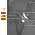
Avulsion fractures of the SPR are only noted on AP and possibly oblique radiographs of the ankle. A vertical avulsion fracture fragment, off the lateral margin of the distal fibula, is nearly pathognomonic for SPR avulsion, with or without associated peroneal tendon dislocation. The SPR avulsion fracture is easily distinguishable from a distal fibular fracture, because it occurs superior and lateral to the fibular tip, with the fragment typically linear in shape, conforming to the lateral fibular cortex ( Figs. 3 and 4 ).


Acute SPR injuries leading to peroneal tendon instability can be readily assessed on axial MR imaging and graded according to the classification first described by Eckert and Davis. According to this classification, the most common injuries include stripping of the SPR from its fibular attachment (see Fig. 4 ), with formation of a subperiosteal pouch into which the peroneal tendons can dislocate, and bony avulsions at the SPR attachment to the fibula. A recent classification, with surgical implications, describes a large lateral fracture fragment, with an intact SPR and dislocation of the peroneal tendons between the fracture fragment and fibula.
Both MR imaging and CT allow for assessment of retro-malleolar groove morphology, because flat, convex, or irregular grooves can predispose to injuries of the SPR and peroneal tendon dislocation. MR imaging also allows for evaluation of concomitant lateral collateral ligament or peroneal tendon tears, whereas periosteal stripping and small avulsion fractures may be better characterized on radiographs or CT (see Fig. 3 ). Given their association with other types of ankle fractures, typically intra-articular calcaneal fractures, SPR injuries may, on occasion, be detected on ankle CT obtained for fracture characterization elsewhere. The SPR can be also be assessed with ultrasound, with the particular advantage of demonstrating transient peroneal tendon subluxation or dislocation with dynamic resisted dorsiflexion-eversion of the ankle.
Conservative management in a short leg cast with the foot in neutral to slight inversion for 6 weeks may be recommended for acute type I and possibly type III injuries. Although several other procedures exist, surgical management most commonly includes anatomic repair of the SPR with fibular groove deepening.
Fracture of the Lateral Process of the Talus
The lateral process of the talus is part of the posterior subtalar and talofibular joints. Although relatively uncommon, fractures of the lateral process of the talus (LTPFs) account for up to 20% of talar fractures. The mechanism of injury is axial loading and dorsiflexion of the foot with external rotation, most commonly associated with snowboarding. LTPFs can also be seen in patients after motor vehicle accidents and falls from height. These fractures clinically present as persistent lateral ankle pain and can be initially misdiagnosed as lateral ankle sprain, with secondary significant morbidity and subtalar arthritis.
Fractures of the lateral process of the talus are frequently detected only on AP radiographs of the ankle, manifesting as inframalleolar bony protuberances and irregularities of the lateral talus ( Fig. 5 ). Secondary subtalar arthritis may depict additional changes in the opposing calcaneus. LTPFs, although difficult to detect, may be noted on the lateral ankle view using the V sign, which is an interruption or asymmetry of the normal V-shaped contour of the lateral process of the talus ( Fig. 6 ). Recent literature, however, suggests that the presence of the V sign varies with the extent of the fracture and foot position, and is subject to interobserver variability. CT is usually required for confirmation of LTPFs in suspected cases and for further characterization of the fracture. Given the often delayed clinical presentation or misdiagnosis as lateral ankle sprain, LTPFs may be seen subacutely or even remotely on MR imaging with evidence of secondary posterior subtalar arthritis (see Fig. 5 ).


Small or nondisplaced LTPFs can be managed nonoperatively with immobilization and progressive weight-bearing, whereas larger and displaced fractures require fixation with lag screws or a miniplate. Significantly comminuted fractures are treated with excision. Nonunited fractures with a late presentation can be excised if small and internally fixed if large. In cases that have progressed to malunion or nonunion, pseudoarthrosis and even lateral hindfoot impingement have been reported.
Fracture of the Anterior Process of the Calcaneus
The anterior process of the calcaneus projects distal to the calcaneal sulcus, articulating with both the talus, via the middle and anterior subtalar joints, and with the cuboid.
Fractures of the anterior process of the calcaneus (APCFs) occur as a result of 2 proposed mechanisms of injury. The more common mechanism involves avulsion fractures of the dorsal calcaneocuboid ligament, the bifurcate ligament, and/or the extensor digitorum brevis muscle during inversion of a plantarflexed ankle. A less common mechanism is shearing/compression during forced dorsiflexion and eversion (nutcracker fracture). Clinically, APCFs present as ecchymosis and tenderness approximately 2 cm distal and 1 cm inferior to the anterior talofibular ligament. As the fractures often coexist with lateral collateral ligament tears, the injury is often mistaken for ankle sprain, with a delayed diagnosis of an average of 22 weeks.
On radiographs, APCFs may only be detected on AP radiographs of the ankle, particularly if radiographs of the foot are not available. Depending on the size of the fracture fragment they are often discerned on lateral and oblique views of the foot or a lateral view of the ankle ( Fig. 7 ). On AP view of the ankle, APCFs are detected by presence of a subfibular bone fragment. In the setting of ankle inversion injury, soft tissue swelling, distal, rather than at the level of the fibula, can be a clue to the presence of this fracture (see Fig. 7 ). Smaller APCFs are thought to be due to avulsion of the dorsal calcaneocuboid and/or bifurcate ligaments, whereas larger avulsion fractures may be due to avulsion at the origin of the extensor digitorum brevis muscle.

APCFs often reflect a component of Chopart joint disruption, or midtarsal sprain, with additional fractures at the talonavicular joints ( Fig. 8 ). Thus, whereas inversion-related distraction forces at the calcaneocuboid joint produce avulsion fractures, typically of the dorsal calcaneocuboid ligament and bifurcate ligament, impaction forces can result in fractures of the navicular and talar head. Concomitant avulsion of the dorsal talonavicular ligament is secondary to simultaneous foot plantarflexion. Therefore, in the setting of APCFs, additional fractures should be scrutinized for, because they indicate a more significant injury of the Chopart joint.

CT and MR imaging are helpful when the APCFs are radiographically occult or nondisplaced, by further characterizing fracture morphology, displacement, and intra-articular extension. The fractures are best visualized in the sagittal plane and are most often vertical in orientation.
MR imaging is useful also for detecting concurrent injuries to the lateral collateral ligament complex, the medial aspect of the Chopart joint, and for assessing the integrity of the dorsal talonavicular, dorsal calcalcaneocuboid, and bifurcate ligaments.
Minimally displaced APCFs can be treated conservatively but may require longer immobilization and nonweight-bearing when compared with ankle sprains. In patients with displaced fractures or with refractory pain, open reduction internal fixation of larger fragments and surgical excision of smaller fragments have both shown satisfactory outcomes.
Enlarged Peroneal Tubercle
The PT, also known as the trochlear process, is an osseous protuberance, located along the lateral calcaneal wall, with commonly reported prevalence of approximately 40%, although higher prevalence of up to 97% has been reported. The PT serves as the attachment site of the inferior peroneal retinaculum, divides the peroneus longus and brevis tendon sheaths, and acts as a pulley or fulcrum for the peroneus longus tendon as it turns toward the cuboid. The peroneus brevis and longus tendons course superior/anterior and inferior/posterior to the PT, respectively.
The mean width of the PT is 3 mm, with 5 mm often used as a cutoff to diagnose an enlarged PT. The reported prevalence of an enlarged PT is approximately 21%. Morphologic variations of the PT have also been described. An enlarged PT displaces the peroneus longus tendon posterolaterally and effectively increases the acuity of the angle that the tendon must take toward the cuboid, predisposing it to increased tension and secondary disease such as tendinosis and tearing. Peroneus brevis disease is less common in the setting of hypertrophied PT.
The PT may only be detected on AP view of the ankle, if no foot films are available, as a lateral osseous protuberance, arising from the calcaneus ( Fig. 9 ). The PT is optimally seen on cross-sectional imaging, with either ultrasonography, CT, or MR imaging of the ankle. On MR imaging, an enlarged PT may demonstrate reactive marrow edema with adjacent tendinosis, tenosynovitis, and longitudinal split tearing of the peroneus longus tendon, and, less commonly, concomitant involvement of the peroneus brevis tendon (see Figs. 9 and 10 ).