DOUBLE AORTIC
ARCH, AND ABERRANT
SUBCLAVIAN ARTERY
 INTRODUCTION
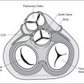
It is generally acknowledged that anomalies of aortic arch branching are best understood when basic embryology of the aortic arch is reviewed. The hypothetical double aortic arch theory, suggested by Edwards (1), provides an explanation for various aortic arch abnormalities (2–4). This theory is based on the presence of double aortic arch in the embryo, where the ascending aorta splits into a right and a left aortic arch, which merge to form the descending aorta, anatomically located in a central position, anterior to the spine (Fig. 21-1). A complete vascular ring, surrounding the trachea and esophagus, is thus formed by the left and right arches. The left and right aortic arches give rise to two vessels each; the left and right common carotid and subclavian arteries, respectively (Fig. 21-1). In addition, the left and right pulmonary arteries are connected to the left and right aortic arch, respectively, by a left and right ductus arteriosus in the region of the subclavian arteries (Fig. 21-1). Normal and abnormal development of the aortic arch branching is thus related to which site of the left or right aortic arch regresses or persists during embryonic development (3,4). Common aortic arch abnormalities encountered on prenatal ultrasound are discussed in this chapter.
 EMBRYOLOGIC FINDINGS
Related posts:


Stay updated, free articles. Join our Telegram channel

Full access? Get Clinical Tree


